
















©The Author(s) 2025.
World J Radiol. Sep 28, 2025; 17(9): 110906
Published online Sep 28, 2025. doi: 10.4329/wjr.v17.i9.110906
Published online Sep 28, 2025. doi: 10.4329/wjr.v17.i9.110906
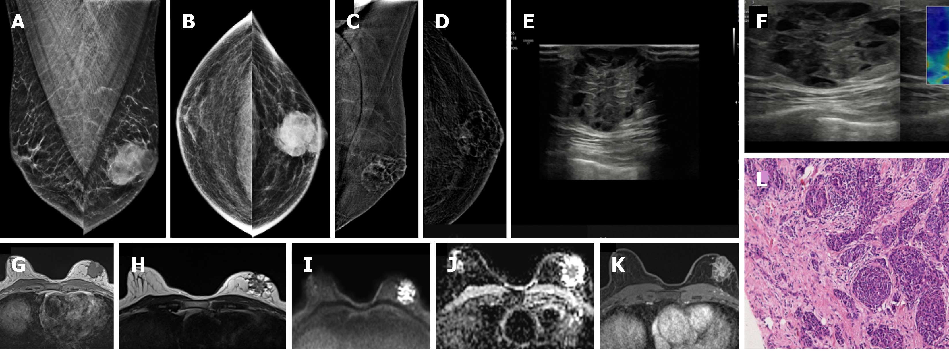
Figure 19 Invasive breast cancer in a 74-year-old male with a hard lump in left breast.
A: Mediolateral oblique (MLO) view; B: Craniocaudal (CC) view showing an irregular, high-density mass; C and D: Contrast-enhanced mammography; C: MLO and D: CC views of the recombined images showing heterogeneous enhancement; E: Ultrasound; F: Elastography images showing an irregular heteroechoic mass with microlobulated margins that appear hard on elastography; G-J: Magnetic resonance images showing an irregular mass; G: T1-weighted image showing a hypointense signal; H: T2-weighted image showing a heterogeneously hyperintense signal; I and J: DWI-ADC images showing central diffusion restriction; K: Postcontrast image showing heterogeneous septal enhancement; L: Histopathological examination image (hematoxylin–eosin, × 40) showing invasive breast cancer (no special type) composed of tumor cells arranged in clusters and tubules.
- Citation: Singla V, Bhatia H, Garg D, Bal A, Sekar A. A compendium of male breast imaging: The road less traveled. World J Radiol 2025; 17(9): 110906
- URL: https://www.wjgnet.com/1949-8470/full/v17/i9/110906.htm
- DOI: https://dx.doi.org/10.4329/wjr.v17.i9.110906