
















©The Author(s) 2025.
World J Radiol. Sep 28, 2025; 17(9): 110906
Published online Sep 28, 2025. doi: 10.4329/wjr.v17.i9.110906
Published online Sep 28, 2025. doi: 10.4329/wjr.v17.i9.110906
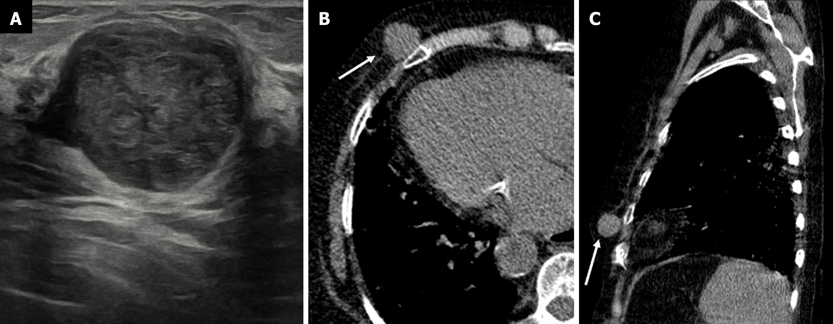
Figure 16 Myofibroblastoma in a 56-year-old male with a palpable mass in the right breast.
A: Ultrasound image showing a round circumscribed heterogeneously hypoechoic mass; B: Axial; C: Sagittal noncontrast computed tomography sections demonstrating a well-defined hypoattenuating soft tissue density mass in the right breast (white arrows) with no calcifications or chest wall invasion.
- Citation: Singla V, Bhatia H, Garg D, Bal A, Sekar A. A compendium of male breast imaging: The road less traveled. World J Radiol 2025; 17(9): 110906
- URL: https://www.wjgnet.com/1949-8470/full/v17/i9/110906.htm
- DOI: https://dx.doi.org/10.4329/wjr.v17.i9.110906