
















©The Author(s) 2025.
World J Radiol. Sep 28, 2025; 17(9): 110906
Published online Sep 28, 2025. doi: 10.4329/wjr.v17.i9.110906
Published online Sep 28, 2025. doi: 10.4329/wjr.v17.i9.110906
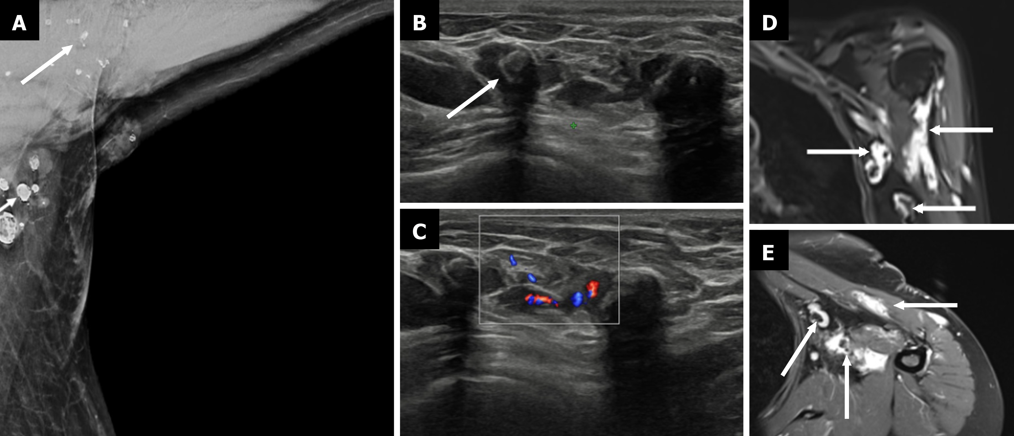
Figure 12 Venolymphatic malformation.
A: Mediolateral oblique mammogram with an axillary view shows multiple well-defined, lobulated densities in the axilla (arrow), some containing coarse calcifications; B: Grayscale ultrasound reveals multiple anechoic and hypoechoic compressible tubular channels in the subcutaneous plane with echogenic phleboliths (arrow), which is consistent with dilated venous and lymphatic components; C: Color Doppler ultrasound demonstrates slow flow within some of the vascular channels; D: Coronal T2-weighted fat-saturated magnetic resonance image (MRI) shows hyperintense lobulated masses (arrows) in the axilla; E: Postcontrast axial T1-weighted fat-suppressed MRI shows enhancement of the lesion (thin arrows), with nonenhancing phleboliths, which is consistent with a benign low-flow vascular malformation.
- Citation: Singla V, Bhatia H, Garg D, Bal A, Sekar A. A compendium of male breast imaging: The road less traveled. World J Radiol 2025; 17(9): 110906
- URL: https://www.wjgnet.com/1949-8470/full/v17/i9/110906.htm
- DOI: https://dx.doi.org/10.4329/wjr.v17.i9.110906