
















©The Author(s) 2025.
World J Radiol. Sep 28, 2025; 17(9): 110906
Published online Sep 28, 2025. doi: 10.4329/wjr.v17.i9.110906
Published online Sep 28, 2025. doi: 10.4329/wjr.v17.i9.110906
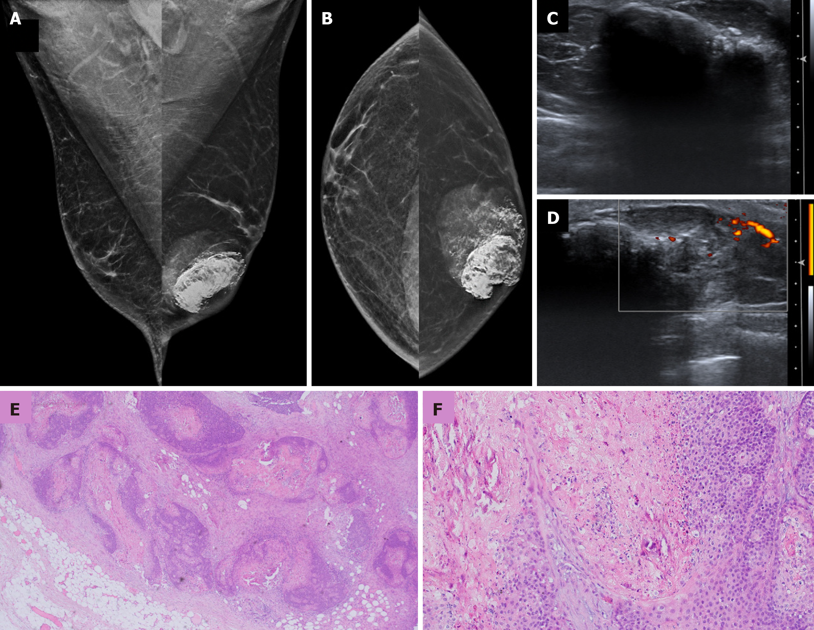
Figure 11 Pilomatricoma in a 33-year-old male with a palpable left breast mass.
A: Mediolateral oblique and B: Craniocaudal mammography images showing a large irregular high-density mass with circumscribed margins and coarse calcifications in the retroareolar location; C: Ultrasound; D: Color Doppler images showing an irregular lesion with extensive posterior acoustic shadowing and some internal vascularity; E and F: Histopathology images (hematoxylin and eosin, × 40) showing a circumscribed lobulated mass in subcutaneous tissue and islands of basaloid cells exhibiting abrupt keratinization without an intervening granular layer along with shadow cells and central calcification.
- Citation: Singla V, Bhatia H, Garg D, Bal A, Sekar A. A compendium of male breast imaging: The road less traveled. World J Radiol 2025; 17(9): 110906
- URL: https://www.wjgnet.com/1949-8470/full/v17/i9/110906.htm
- DOI: https://dx.doi.org/10.4329/wjr.v17.i9.110906