
















©The Author(s) 2025.
World J Radiol. Sep 28, 2025; 17(9): 110267
Published online Sep 28, 2025. doi: 10.4329/wjr.v17.i9.110267
Published online Sep 28, 2025. doi: 10.4329/wjr.v17.i9.110267
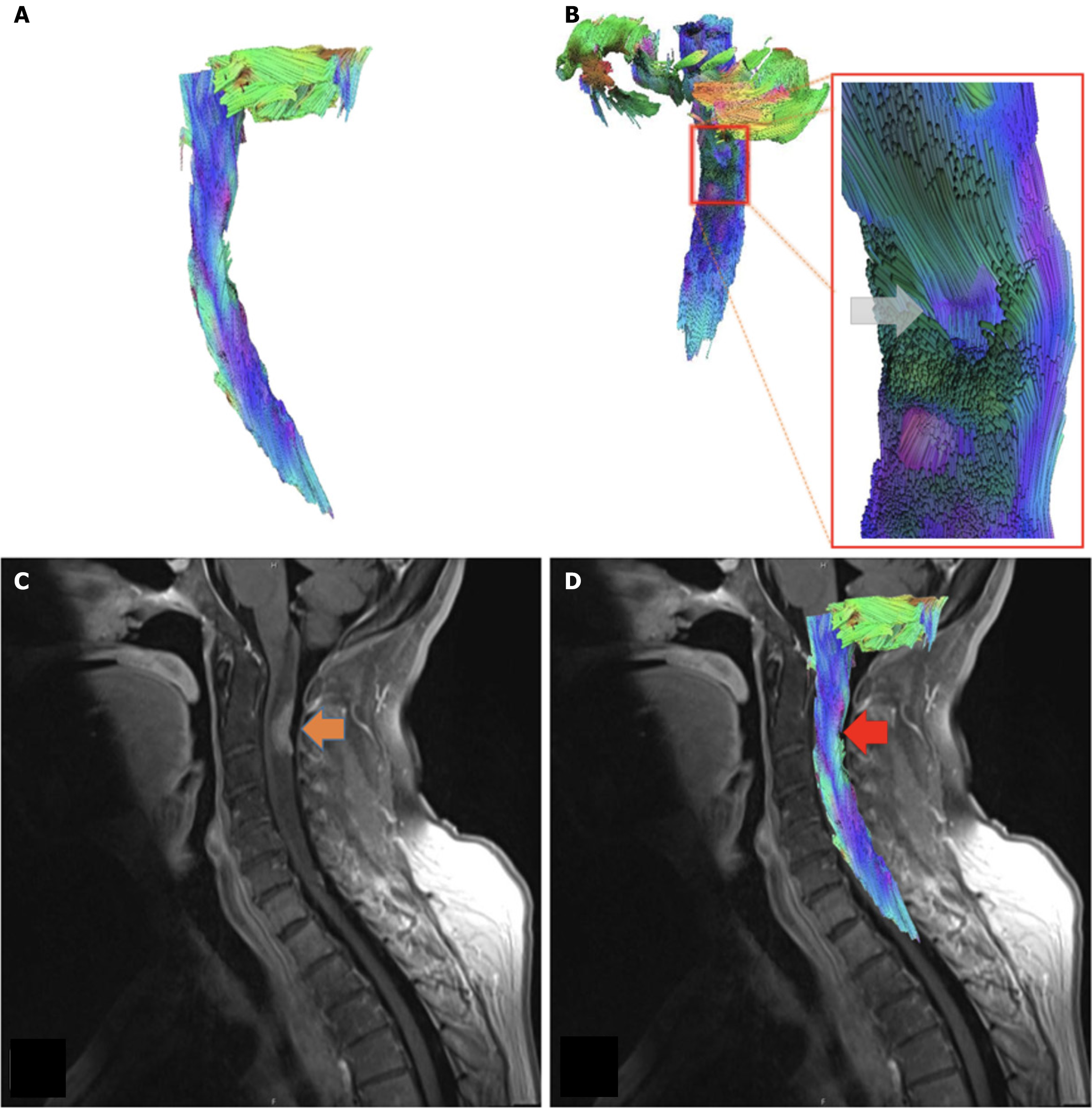
Figure 3 Metastasis case.
A: Sagittal view of tractography model. Case of 62-year-old female with primary breast cancer, displaying metastases to the lungs, spine, and brain; B: Posterior oblique view of the cervical spinal cord tractography model, with associated close up at the level of C2-C3. Note displaced fibers at the site of lesion (gray arrow); C: T1 FR STIR sagittal view of the cervical spinal cord. Metastatic lesion located at C2-C3 (orange arrow); D: Tractography model of metastasis case superimposed over T1 FR STIR sagittal view of the cervical spinal cord. The metastatic lesion can be seen at the level C2-C3 (red arrow).
- Citation: Supsupin EP, Serrano A, Louviere C, Pearson L, Hernandez M, Sekar V, Amer A, Cikla U, Virarkar M, Gumus KZ. Magnetic resonance tractography of the cervical spine: A rapid diffusion tensor imaging protocol to serve as a clinical evaluation tool. World J Radiol 2025; 17(9): 110267
- URL: https://www.wjgnet.com/1949-8470/full/v17/i9/110267.htm
- DOI: https://dx.doi.org/10.4329/wjr.v17.i9.110267