
















©The Author(s) 2025.
World J Radiol. Sep 28, 2025; 17(9): 110267
Published online Sep 28, 2025. doi: 10.4329/wjr.v17.i9.110267
Published online Sep 28, 2025. doi: 10.4329/wjr.v17.i9.110267
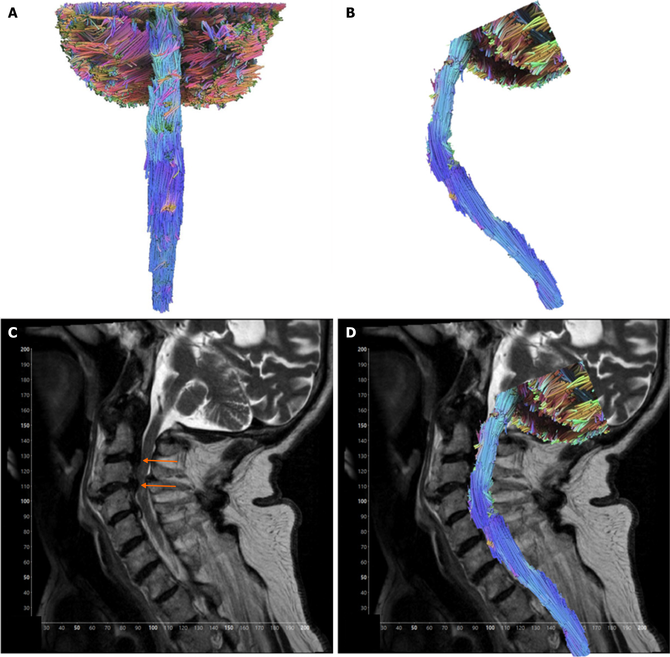
Figure 2 Contusion case.
A: AP view of tractography model. Case of an 82-year-old male presenting with spinal cord contusion; B: Sagittal view of the tractography model; C: T2-weighted sagittal view of the cervical spinal cord showing severe spinal canal stenosis from disc herniation and degenerative spondylosis at the levels of C2-C3 and C3-C4, with severe spinal cord flattening at C3-C4 (orange arrows); D: Sagittal view tractography model of contusion case superimposed over T2-weighted imaging of the cervical spinal cord.
- Citation: Supsupin EP, Serrano A, Louviere C, Pearson L, Hernandez M, Sekar V, Amer A, Cikla U, Virarkar M, Gumus KZ. Magnetic resonance tractography of the cervical spine: A rapid diffusion tensor imaging protocol to serve as a clinical evaluation tool. World J Radiol 2025; 17(9): 110267
- URL: https://www.wjgnet.com/1949-8470/full/v17/i9/110267.htm
- DOI: https://dx.doi.org/10.4329/wjr.v17.i9.110267