
















©The Author(s) 2025.
World J Radiol. Sep 28, 2025; 17(9): 110214
Published online Sep 28, 2025. doi: 10.4329/wjr.v17.i9.110214
Published online Sep 28, 2025. doi: 10.4329/wjr.v17.i9.110214
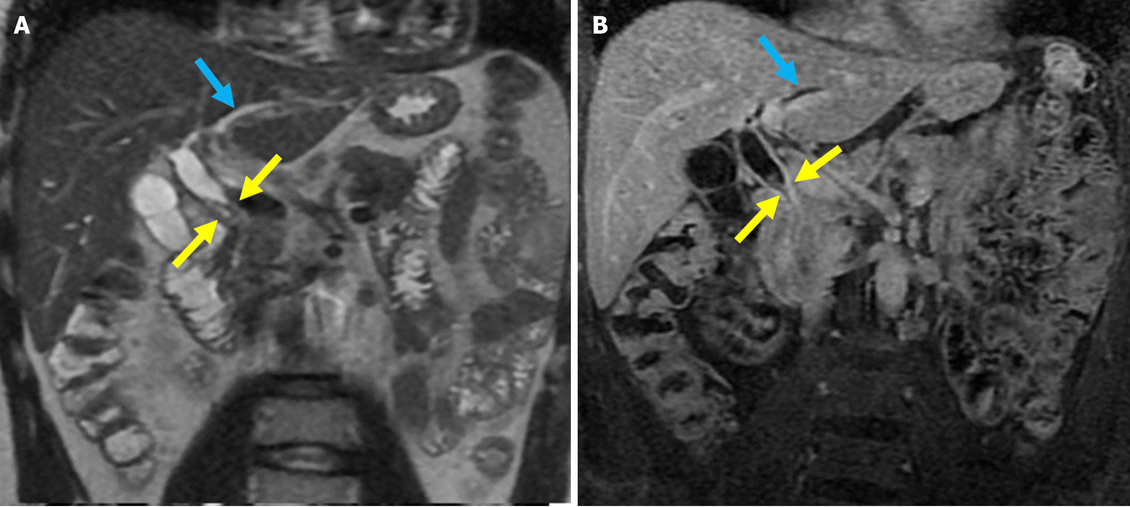
Figure 20 Post-sphincterotomy biliary stricture.
A: Coronal T2-weighted magnetic resonance imaging; B: Fat-suppressed post-contrast T1-weighted magnetic resonance imaging showed a biliary stricture in a 40-year-old male patient with a history of endoscopic retrograde cholangiopancreatography and sphincterotomy. Focal stenosis was observed in the mid portion of the common bile duct without any associated mass lesion (yellow arrows). There was associated mural thickening of the mid-bile duct and dilatation of the intrahepatic bile ducts (blue arrows). The findings were consistent with a benign biliary stricture secondary to prior endoscopic retrograde cholangiopancreatography.
- Citation: Simsar M, Yuruk YY, Sahin O, Sahin H. Complications and expected imaging findings after endoscopic retrograde cholangiopancreatography. World J Radiol 2025; 17(9): 110214
- URL: https://www.wjgnet.com/1949-8470/full/v17/i9/110214.htm
- DOI: https://dx.doi.org/10.4329/wjr.v17.i9.110214