
















©The Author(s) 2025.
World J Radiol. Sep 28, 2025; 17(9): 110214
Published online Sep 28, 2025. doi: 10.4329/wjr.v17.i9.110214
Published online Sep 28, 2025. doi: 10.4329/wjr.v17.i9.110214
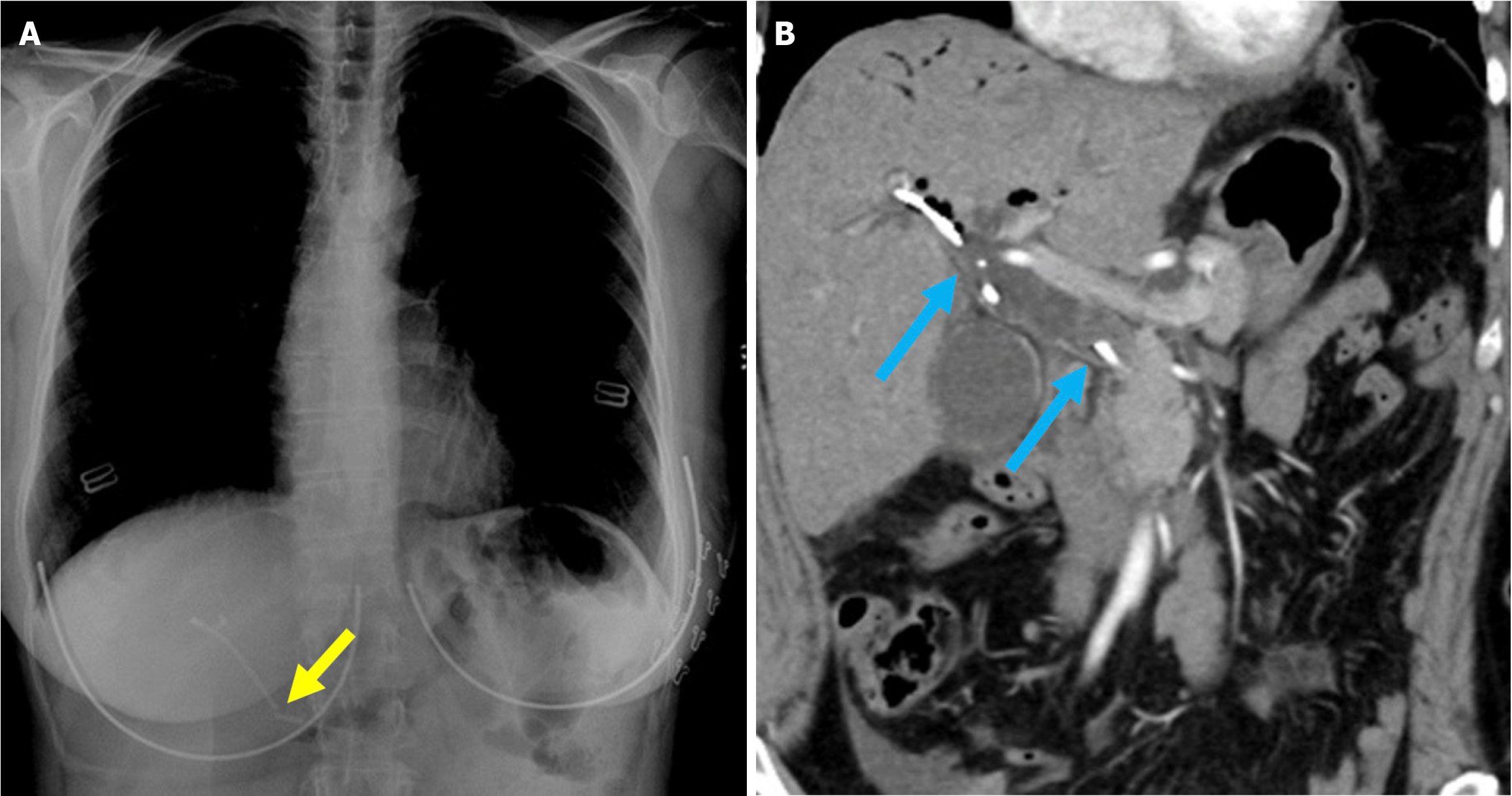
Figure 19 Stent fracture.
A: Posteroanterior chest radiograph; B: Contrast-enhanced coronal computed tomography scan. A 52-year-old female patient presented to the hospital 3 days after biliary stent placement via endoscopic retrograde cholangiopancreatography with complaints of abdominal pain and elevated levels of aspartate aminotransferase, alanine aminotransferase, and bilirubin. The imaging revealed a fracture of the previously placed plastic stent (yellow arrow). A repeat endoscopic retrograde cholangiopancreatography was performed to remove the fractured stent, but residual stent fragments remained within the common bile duct. The duct appeared dilated due to malfunction of the broken stent (blue arrow).
- Citation: Simsar M, Yuruk YY, Sahin O, Sahin H. Complications and expected imaging findings after endoscopic retrograde cholangiopancreatography. World J Radiol 2025; 17(9): 110214
- URL: https://www.wjgnet.com/1949-8470/full/v17/i9/110214.htm
- DOI: https://dx.doi.org/10.4329/wjr.v17.i9.110214