
















©The Author(s) 2025.
World J Radiol. Sep 28, 2025; 17(9): 110214
Published online Sep 28, 2025. doi: 10.4329/wjr.v17.i9.110214
Published online Sep 28, 2025. doi: 10.4329/wjr.v17.i9.110214
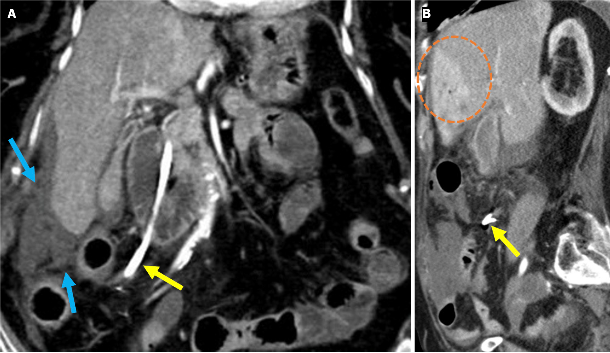
Figure 18 Distal stent migration and duodenal perforation.
A and B: Coronal (A) and sagittal (B) contrast-enhanced computed tomography images in a 77-year-old female patient with a history of endoscopic retrograde cholangiopancreatography and biliary stenting for cholangitis, who presented with severe abdominal pain. Tachypnea and agitation were observed and were accompanied by nausea, fatigue, and impaired oral intake. Laboratory tests revealed elevated leukocyte counts, C-reactive protein levels, and lactate levels. The stent was found to have migrated distally into the duodenum and perforated the distal portion of the second part of the duodenum (yellow arrow). High attenuation-free fluid, suggestive of hemorrhage, was observed in the perihepatic region and mesentery (blue arrows). The liver exhibited heterogeneous contrast enhancement, consistent with impaired perfusion that was likely related to ischemia (orange circle). Emergent surgical intervention was required.
- Citation: Simsar M, Yuruk YY, Sahin O, Sahin H. Complications and expected imaging findings after endoscopic retrograde cholangiopancreatography. World J Radiol 2025; 17(9): 110214
- URL: https://www.wjgnet.com/1949-8470/full/v17/i9/110214.htm
- DOI: https://dx.doi.org/10.4329/wjr.v17.i9.110214