
















©The Author(s) 2025.
World J Radiol. Sep 28, 2025; 17(9): 110214
Published online Sep 28, 2025. doi: 10.4329/wjr.v17.i9.110214
Published online Sep 28, 2025. doi: 10.4329/wjr.v17.i9.110214
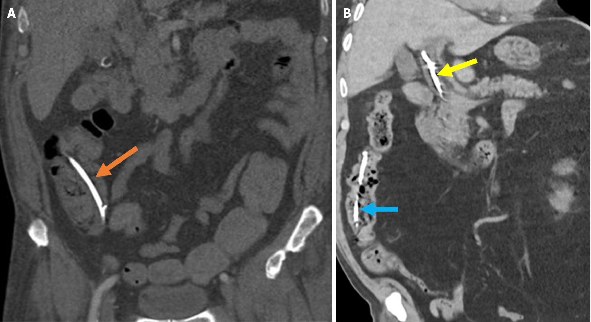
Figure 17 Distal stent migration.
A: A 56-year-old male patient with a history of endoscopic retrograde cholangiopancreatography (ERCP) and biliary stenting presented with right lower quadrant pain lasting for 3 days. The coronal noncontrast computed tomography (CT) images demonstrated distal migration of the stent into the cecum (orange arrow); B: An 85-year-old patient with a history of ERCP and biliary stenting presented with jaundice. The previous stent was not visualized during ERCP for stent revision, and re-stenting was performed (yellow arrow). Post-procedural coronal contrast-enhanced CT revealed distal migration of the previous stent into the ascending colon (blue arrow).
- Citation: Simsar M, Yuruk YY, Sahin O, Sahin H. Complications and expected imaging findings after endoscopic retrograde cholangiopancreatography. World J Radiol 2025; 17(9): 110214
- URL: https://www.wjgnet.com/1949-8470/full/v17/i9/110214.htm
- DOI: https://dx.doi.org/10.4329/wjr.v17.i9.110214