











©The Author(s) 2025.
World J Radiol. Sep 28, 2025; 17(9): 110214
Published online Sep 28, 2025. doi: 10.4329/wjr.v17.i9.110214
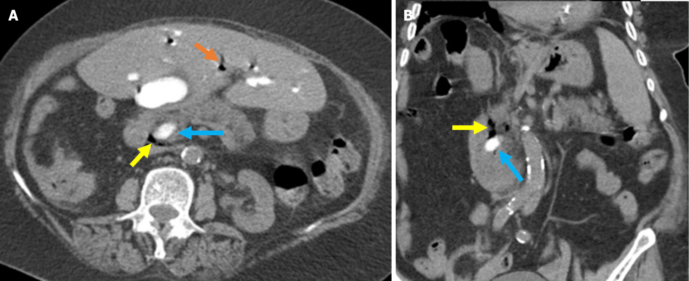
Figure 13 Stapfer type II perforation.
A and B: Axial (A) and coronal (B) noncontrast computed tomography (CT) images in a 70-year-old patient who de veloped a perforation around the ampulla of Vater during sphincterotomy and cannulation. Four hours after the procedure, the patient developed mild epigastric pain, which progressively became diffuse. Eight hours after the procedure, the patient experience fever and tachycardia, which indicated a potential perforation, and a CT scan was subsequently performed. Free air was visible along the medial aspect of the duodenum at the pancreaticoduodenal groove, indicating perforation (yellow arrows). Typical post-endoscopic retrograde cholangiopancreatography appearances included air within the intrahepatic bile ducts (orange arrow) and contrast material within the common bile duct (blue arrows). The patient was managed conservatively.
- Citation: Simsar M, Yuruk YY, Sahin O, Sahin H. Complications and expected imaging findings after endoscopic retrograde cholangiopancreatography. World J Radiol 2025; 17(9): 110214
- URL: https://www.wjgnet.com/1949-8470/full/v17/i9/110214.htm
- DOI: https://dx.doi.org/10.4329/wjr.v17.i9.110214