
















©The Author(s) 2025.
World J Radiol. Sep 28, 2025; 17(9): 110214
Published online Sep 28, 2025. doi: 10.4329/wjr.v17.i9.110214
Published online Sep 28, 2025. doi: 10.4329/wjr.v17.i9.110214
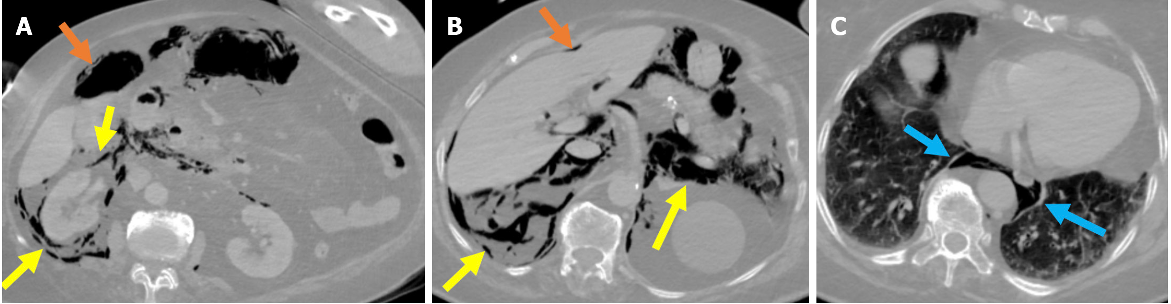
Figure 12 Stapfer type I perforation.
A-C: Contrast-enhanced computed tomography (CT) images in lung window settings at the duodenum (A), celiac artery (B), and basal thorax (C) revealed a lateral duodenal wall perforation in an 84-year-old female patient who had undergone sphincterotomy. During the procedure, suspicion of perforation arose due to suboptimal luminal insufflation during the endoscopic manipulation. A prompt CT scan was performed before the patient was returned to the ward. Findings included pneumoperitoneum (orange arrows) in the perihepatic and right paracolic regions and extensive retroperitoneal emphysema (yellow arrows) involving the right perirenal space, posterior and anterior pararenal spaces, and posterior pneumomediastinum (blue arrows). Due to worsening clinical signs of peritonitis and the results from imaging, an emergency laparotomy was required.
- Citation: Simsar M, Yuruk YY, Sahin O, Sahin H. Complications and expected imaging findings after endoscopic retrograde cholangiopancreatography. World J Radiol 2025; 17(9): 110214
- URL: https://www.wjgnet.com/1949-8470/full/v17/i9/110214.htm
- DOI: https://dx.doi.org/10.4329/wjr.v17.i9.110214