
















©The Author(s) 2025.
World J Radiol. Sep 28, 2025; 17(9): 110214
Published online Sep 28, 2025. doi: 10.4329/wjr.v17.i9.110214
Published online Sep 28, 2025. doi: 10.4329/wjr.v17.i9.110214
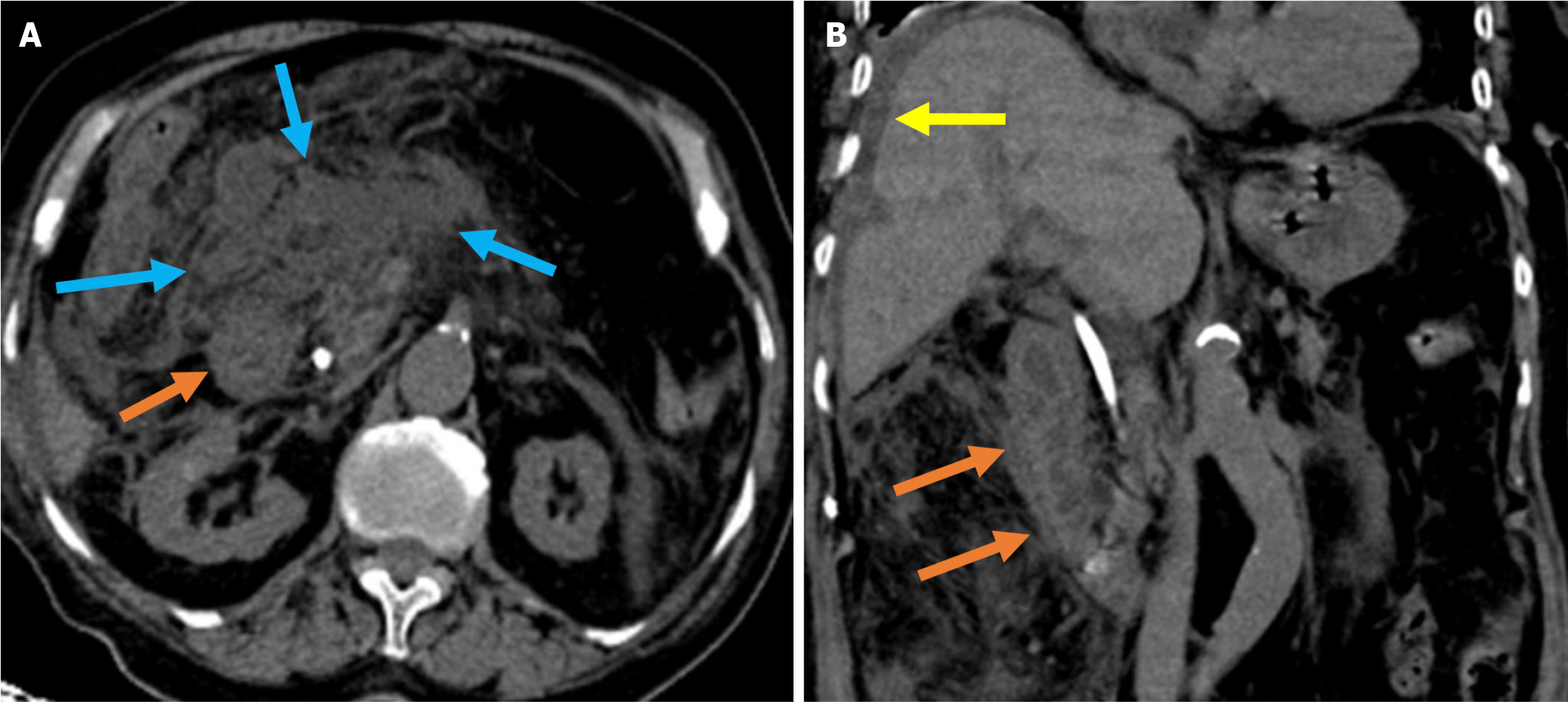
Figure 9 Acute duodenal wall and mesenteric hemorrhages.
A and B: Axial (A) and coronal (B) noncontrast computed tomography images revealed findings consistent with hemorrhage in an 81-year-old female patient who presented with abdominal pain and a drop in hemoglobin levels following endoscopic retrograde cholangiopancreatography. A high-density mesenteric collection and mesenteric fat stranding were observed and consistent with mesenteric hematoma (blue arrows). Hyperattenuating duodenal wall thickening, indicative of an intramural hematoma (orange arrows), was also present. Free fluid was noted in the perihepatic space (yellow arrow).
- Citation: Simsar M, Yuruk YY, Sahin O, Sahin H. Complications and expected imaging findings after endoscopic retrograde cholangiopancreatography. World J Radiol 2025; 17(9): 110214
- URL: https://www.wjgnet.com/1949-8470/full/v17/i9/110214.htm
- DOI: https://dx.doi.org/10.4329/wjr.v17.i9.110214