
















©The Author(s) 2025.
World J Radiol. Sep 28, 2025; 17(9): 110214
Published online Sep 28, 2025. doi: 10.4329/wjr.v17.i9.110214
Published online Sep 28, 2025. doi: 10.4329/wjr.v17.i9.110214
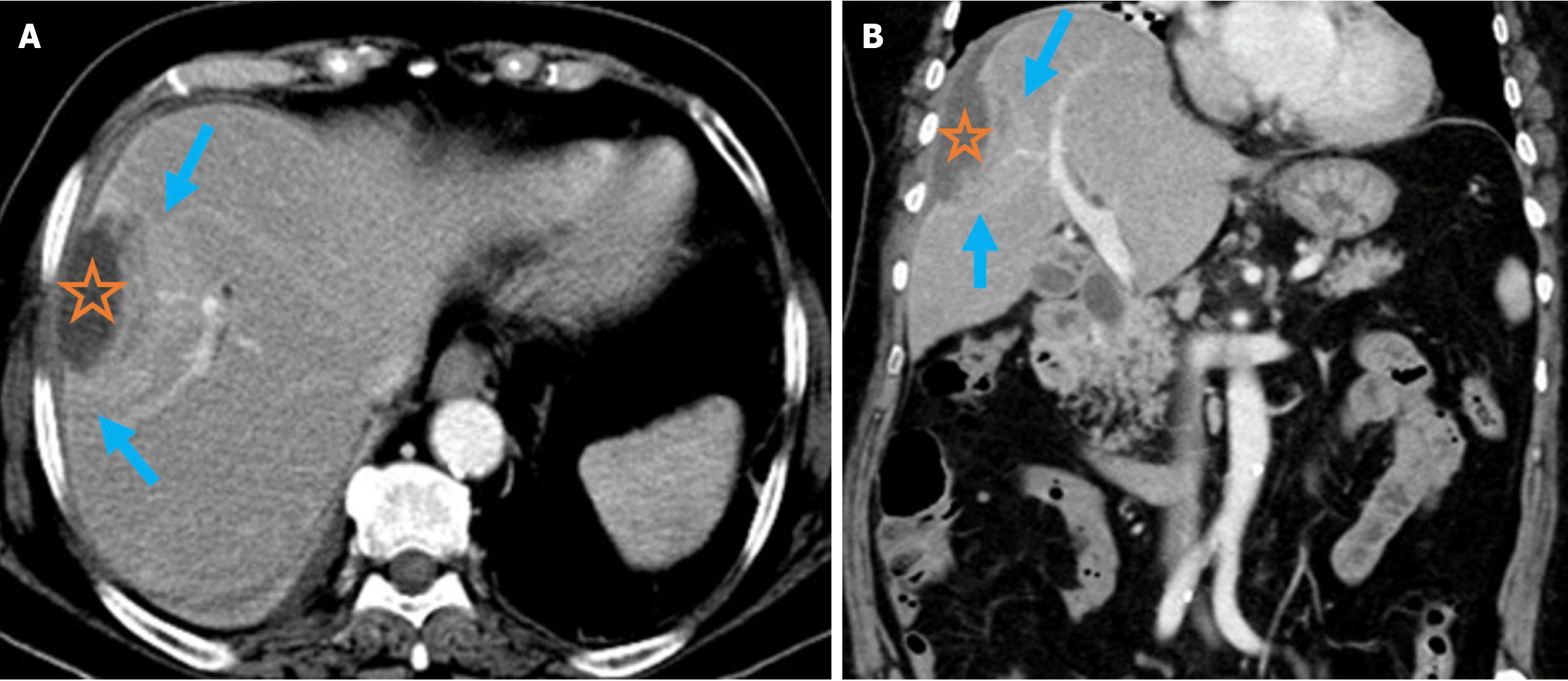
Figure 8 Pyogenic liver abscess.
A and B: Axial (A) and coronal (B) contrast-enhanced computed tomography images demonstrated pyogenic liver abscess secondary to endoscopic retrograde cholangiopancreatography in a 76-year-old female patient. She presented 2 months after endoscopic retrograde cho
- Citation: Simsar M, Yuruk YY, Sahin O, Sahin H. Complications and expected imaging findings after endoscopic retrograde cholangiopancreatography. World J Radiol 2025; 17(9): 110214
- URL: https://www.wjgnet.com/1949-8470/full/v17/i9/110214.htm
- DOI: https://dx.doi.org/10.4329/wjr.v17.i9.110214