
















©The Author(s) 2025.
World J Radiol. Sep 28, 2025; 17(9): 110214
Published online Sep 28, 2025. doi: 10.4329/wjr.v17.i9.110214
Published online Sep 28, 2025. doi: 10.4329/wjr.v17.i9.110214
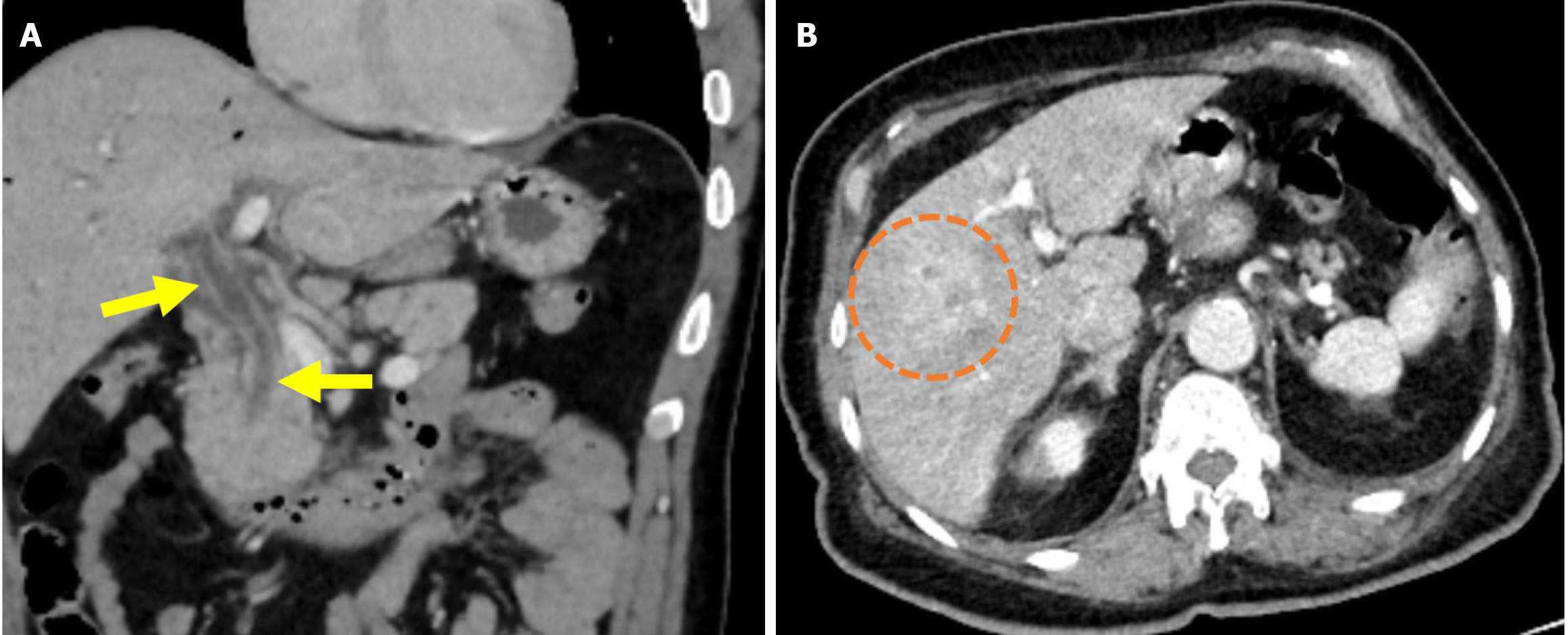
Figure 6 Cholangitis.
A and B: Coronal (A) and axial (B) contrast-enhanced computed tomography images showed findings in a 55-year-old male patient who presented with abdominal pain, chills, and jaundice 1 week after endoscopic retrograde cholangiopancreatography. The patient’s laboratory results revealed elevated levels of gamma-glutamyl transferase, alkaline phosphatase, aspartate aminotransferase, alanine aminotransferase, bilirubin, and acute phase reactants. The patient was subsequently diagnosed with cholangitis. The common bile duct appeared dilated with thickened and enhanced walls (yellow arrows). There was also dilatation of the intrahepatic bile ducts and periductal areas of increased perfusion, likely secondary to inflammation (orange circle).
- Citation: Simsar M, Yuruk YY, Sahin O, Sahin H. Complications and expected imaging findings after endoscopic retrograde cholangiopancreatography. World J Radiol 2025; 17(9): 110214
- URL: https://www.wjgnet.com/1949-8470/full/v17/i9/110214.htm
- DOI: https://dx.doi.org/10.4329/wjr.v17.i9.110214