
















©The Author(s) 2025.
World J Radiol. Sep 28, 2025; 17(9): 110214
Published online Sep 28, 2025. doi: 10.4329/wjr.v17.i9.110214
Published online Sep 28, 2025. doi: 10.4329/wjr.v17.i9.110214
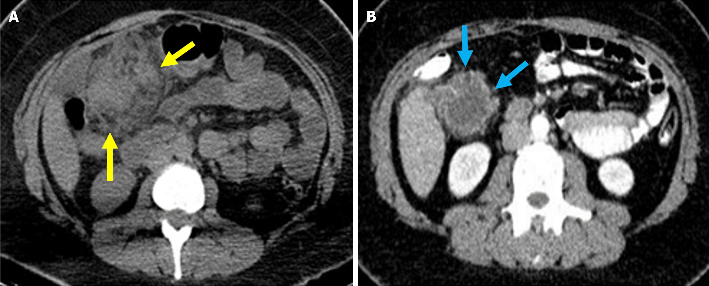
Figure 5 Acute peripancreatic fluid collections and pseudocyst.
A: Noncontrast axial computed tomography (CT); B: Contrast-enhanced axial CT after 15 months. A 28-year-old female patient underwent endoscopic retrograde cholangiopancreatography for choledocholithiasis. Ten hours after the procedure, the patient developed severe upper quadrant abdominal pain accompanied by fever. No signs of peritonitis were observed, and there was no evidence of hemodynamic instability. Laboratory tests revealed elevated levels of pancreatic enzymes and inflammatory markers. Due to suspected pancreatitis a noncontrast CT performed on post-procedure day 1 (A) demonstrated fluid loculation in the infrahepatic and paraduodenal region that was consistent with acute peripancreatic fluid collections (yellow arrow). The patient was diagnosed with post-endoscopic retrograde cholangiopancreatography edematous pancreatitis and was managed conservatively. A follow-up contrast-enhanced CT performed 1.5 months later (B) showed fluid collection in the exact location with pseudocapsule formation that was consistent with a pseudocyst (blue arrow).
- Citation: Simsar M, Yuruk YY, Sahin O, Sahin H. Complications and expected imaging findings after endoscopic retrograde cholangiopancreatography. World J Radiol 2025; 17(9): 110214
- URL: https://www.wjgnet.com/1949-8470/full/v17/i9/110214.htm
- DOI: https://dx.doi.org/10.4329/wjr.v17.i9.110214