
















©The Author(s) 2025.
World J Radiol. Sep 28, 2025; 17(9): 110214
Published online Sep 28, 2025. doi: 10.4329/wjr.v17.i9.110214
Published online Sep 28, 2025. doi: 10.4329/wjr.v17.i9.110214
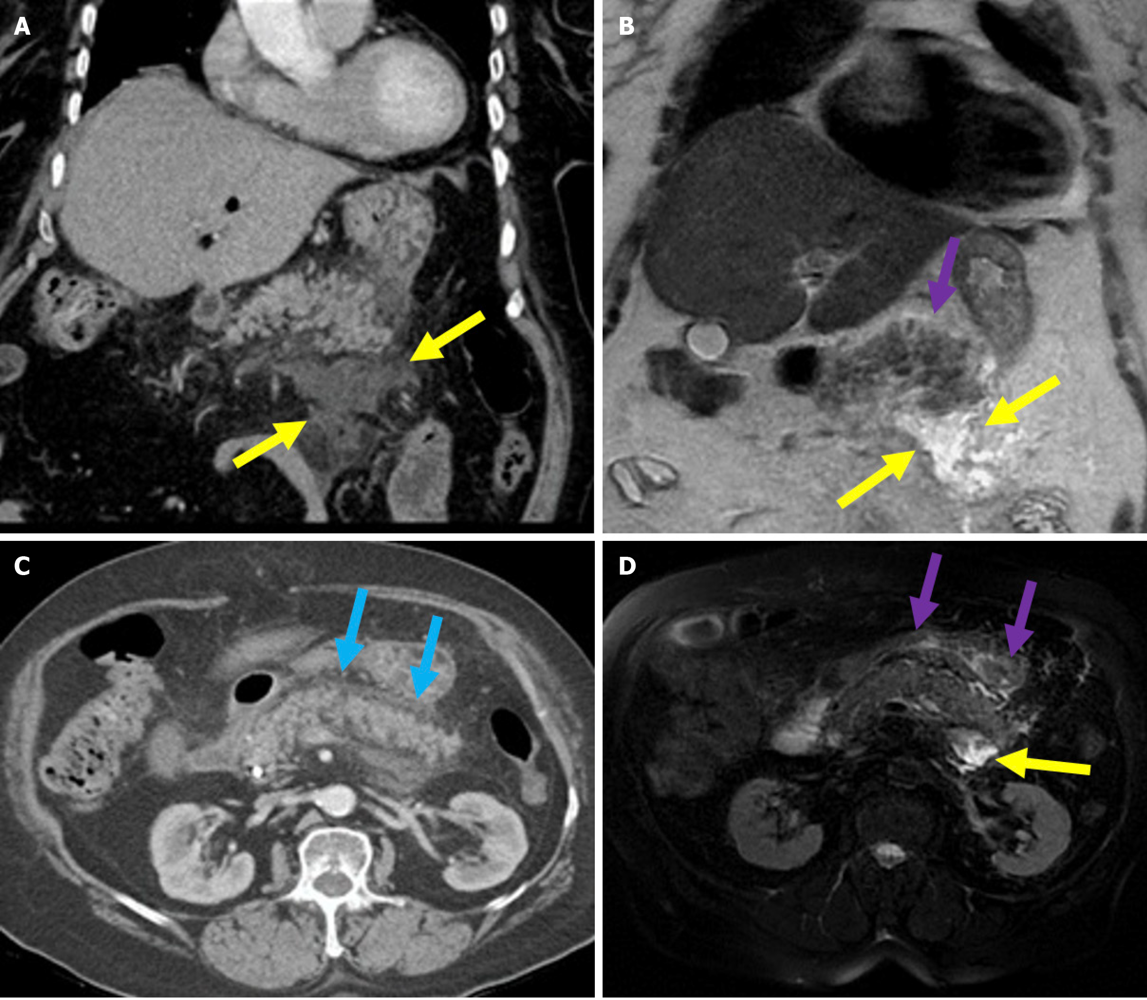
Figure 4 Interstitial edematous pancreatitis and acute peripancreatic fluid collections.
A-C: Contrast-enhanced coronal and axial computed tomography (CT) images (A and C), coronal T2-weighted magnetic resonance imaging (B); D: Fat-suppressed axial T2-weighted magnetic resonance imaging. The 81-year-old female patient diagnosed with post-endoscopic retrograde cholangiopancreatography pancreatitis had presented with epigastric pain and elevated amylase and C-reactive protein levels. CT images showed preserved pancreatic enhancement that was consistent with the absence of necrosis and peripancreatic fat stranding (blue arrow). The pancreatic enlargement and increased T2 signal intensity were suggestive of edema (purple arrow). The pancreas also exhibited mildly diffuse, lace-like T2 hyperintensity within the parenchyma that was associated with a small acute peripancreatic fluid collection (yellow arrow).
- Citation: Simsar M, Yuruk YY, Sahin O, Sahin H. Complications and expected imaging findings after endoscopic retrograde cholangiopancreatography. World J Radiol 2025; 17(9): 110214
- URL: https://www.wjgnet.com/1949-8470/full/v17/i9/110214.htm
- DOI: https://dx.doi.org/10.4329/wjr.v17.i9.110214