















©The Author(s) 2025.
World J Radiol. Sep 28, 2025; 17(9): 110214
Published online Sep 28, 2025. doi: 10.4329/wjr.v17.i9.110214
Published online Sep 28, 2025. doi: 10.4329/wjr.v17.i9.110214
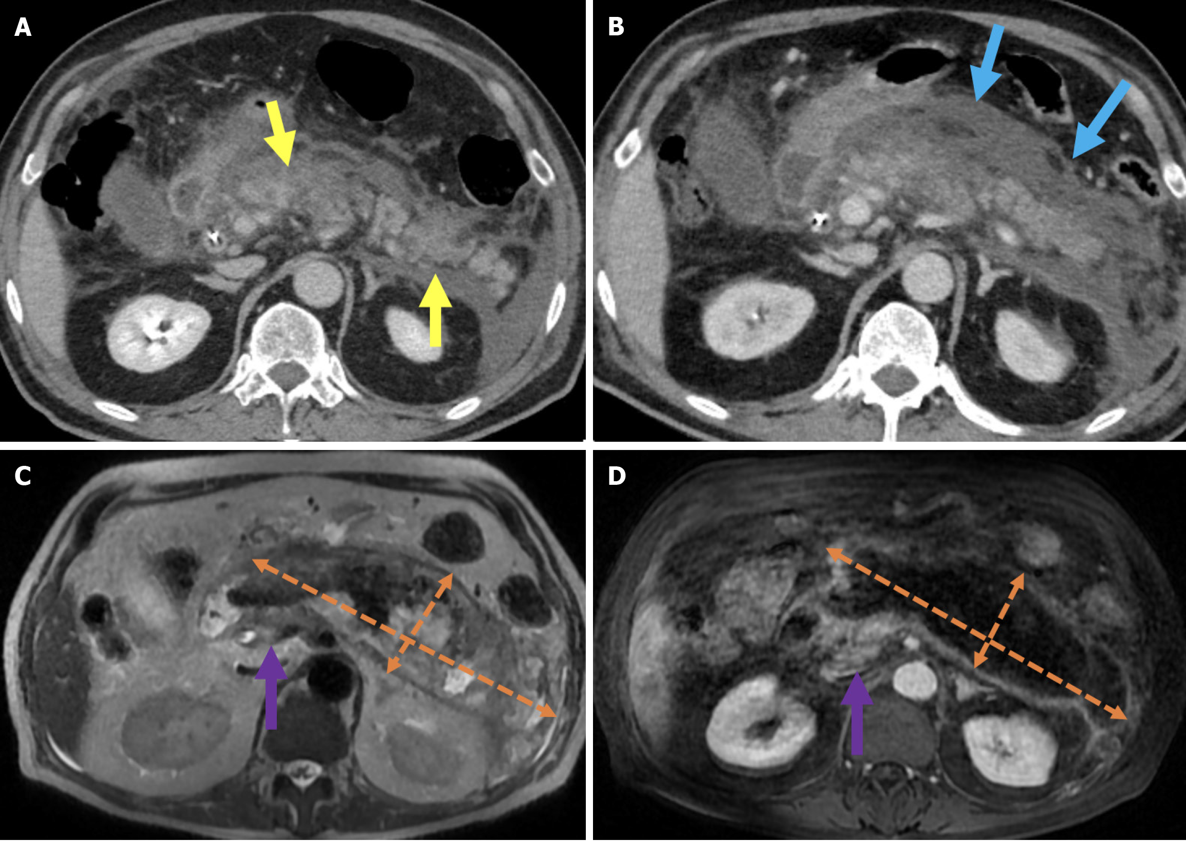
Figure 3 Necrotizing pancreatitis, acute necrotic collections, and walled-off necrosis.
A and B: Contrast-enhanced axial computed tomography (CT); C: T2-weighted magnetic resonance imaging (MRI); D: Post-contrast fat-suppressed T1-weighted MRI. A 76-year-old male patient underwent endoscopic retrograde cholangiopancreatography due to choledocholithiasis. On the evening of the procedure, the patient developed acute abdominal pain that was unresponsive to analgesics. Laboratory tests revealed elevated levels of aspartate aminotransferase, alanine aminotransferase, and lipase. Within the following 24 hours, the patient’s condition deteriorated and developed into shock. Subsequently, the patient required vasopressor support and intubation. Contrast-enhanced CT (A and B) was performed due to the severe abdominal pain. The images showed a markedly enlarged pancreas with areas of non-enhancement consistent with necrotizing pancreatitis that was accompanied by peripancreatic edema (yellow arrows). Heterogeneous acute necrotic collections were also observed in the peripancreatic region (blue arrows). Two months later, an MRI was performed for follow-up. On T2-weighted images (C) a collection with a heterogeneous internal structure and defined wall that was consistent with walled-off necrosis was visualized (orange double-sided arrow). There was a significant loss of pancreatic volume due to necrosis. In the post-contrast T1-weighted fat-suppressed image (D), contrast enhancement of the wall of the collection was noted. A small remnant of pancreatic tissue with normal contrast enhancement was observed (purple arrow).
- Citation: Simsar M, Yuruk YY, Sahin O, Sahin H. Complications and expected imaging findings after endoscopic retrograde cholangiopancreatography. World J Radiol 2025; 17(9): 110214
- URL: https://www.wjgnet.com/1949-8470/full/v17/i9/110214.htm
- DOI: https://dx.doi.org/10.4329/wjr.v17.i9.110214