
















©The Author(s) 2025.
World J Radiol. Sep 28, 2025; 17(9): 110214
Published online Sep 28, 2025. doi: 10.4329/wjr.v17.i9.110214
Published online Sep 28, 2025. doi: 10.4329/wjr.v17.i9.110214
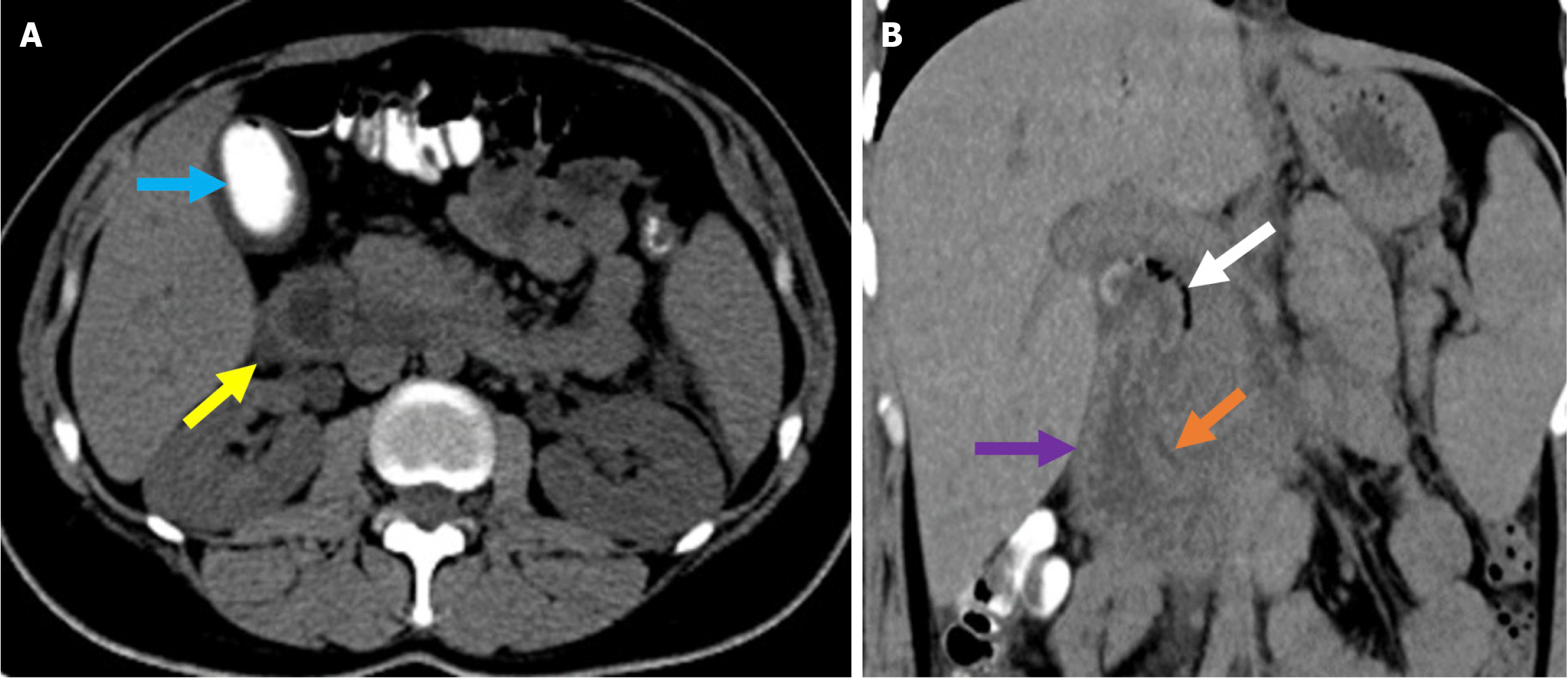
Figure 2 Typical imaging finding of acute duodenitis after endoscopic retrograde cholangiopancreatography.
A and B: Axial (A) and coronal (B) noncontrast computed tomography images revealed post-endoscopic retrograde cholangiopancreatography acute reversible duodenitis in a 26-year-old female patient. Following sphincterotomy, epigastric pain and dyspeptic symptoms developed the next day. Due to the persistence of symptoms during follow-up, imaging was performed. Periduodenal fat stranding (yellow arrow) and increased thickness with submucosal edema of the duodenal wall (purple arrow) were observed. Minimal fluid was noted in the pancreaticoduodenal groove (orange arrow). Expected post-procedural findings include intraductal air within the biliary tree (white arrow) and contrast material within the gallbladder (blue arrow).
- Citation: Simsar M, Yuruk YY, Sahin O, Sahin H. Complications and expected imaging findings after endoscopic retrograde cholangiopancreatography. World J Radiol 2025; 17(9): 110214
- URL: https://www.wjgnet.com/1949-8470/full/v17/i9/110214.htm
- DOI: https://dx.doi.org/10.4329/wjr.v17.i9.110214