Copyright
©The Author(s) 2025.
World J Radiol. Aug 28, 2025; 17(8): 109447
Published online Aug 28, 2025. doi: 10.4329/wjr.v17.i8.109447
Published online Aug 28, 2025. doi: 10.4329/wjr.v17.i8.109447
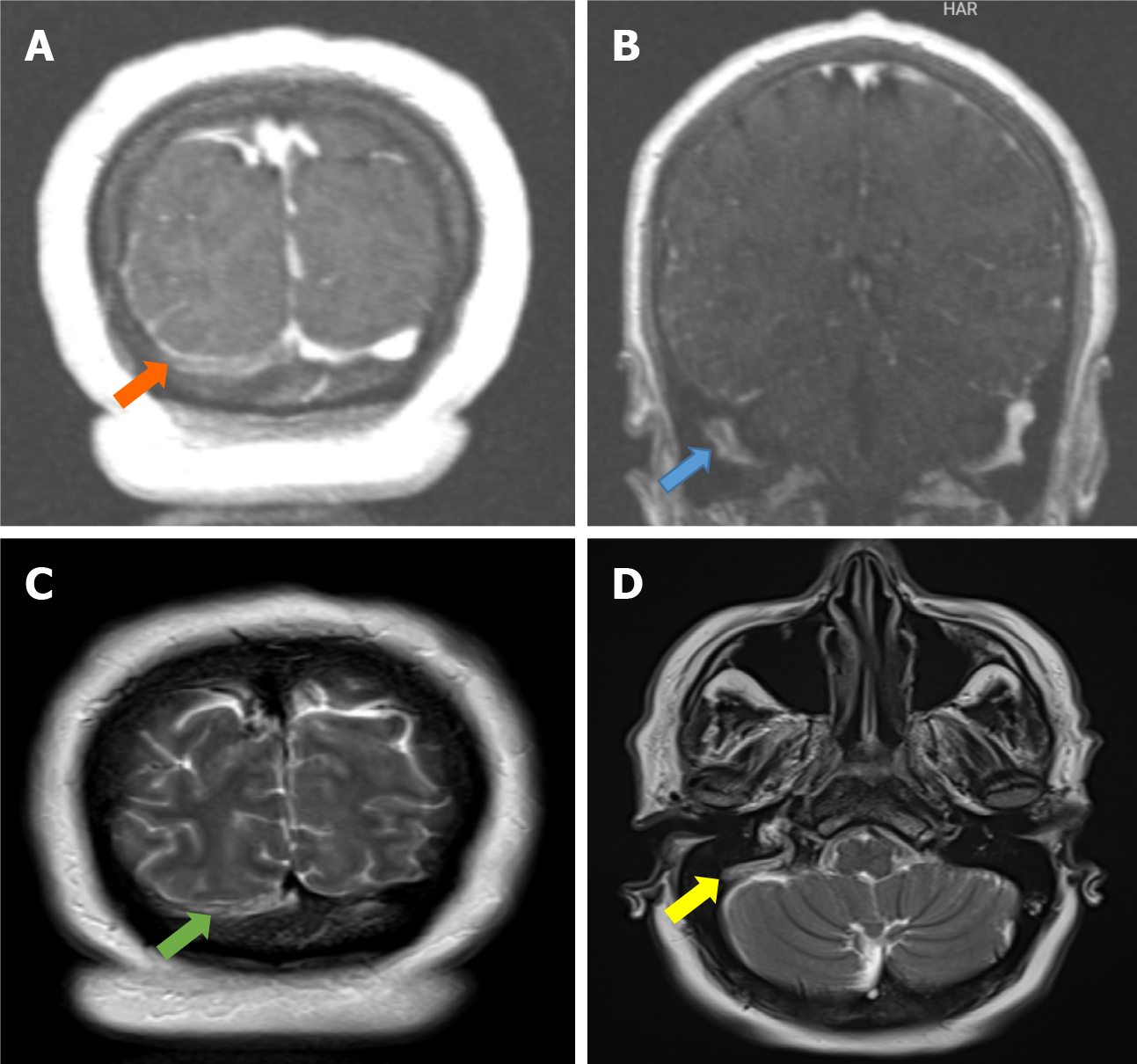
Figure 4 Lateral sinus thrombosis.
A: The coronal post-contrast T1W cerebral magnetic resonance venography image shows loss of signal along the right transverse sinus (orange arrow); B: The coronal post-contrast T1W cerebral magnetic resonance venography image shows loss of signal along the right sigmoid sinus (blue arrow);C: The coronal T2W magnetic resonance image shows hyperintensity due to flow void loss in the right transverse sinus (green arrow); D: Axial T2W magnetic resonance image shows hyperintensity due to flow void loss in the right sigmoid sinus (yellow arrow).
- Citation: Memis KB, Aydin S. Role of imaging in chronic otitis media and its complications. World J Radiol 2025; 17(8): 109447
- URL: https://www.wjgnet.com/1949-8470/full/v17/i8/109447.htm
- DOI: https://dx.doi.org/10.4329/wjr.v17.i8.109447


