
















©The Author(s) 2025.
World J Radiol. Aug 28, 2025; 17(8): 109447
Published online Aug 28, 2025. doi: 10.4329/wjr.v17.i8.109447
Published online Aug 28, 2025. doi: 10.4329/wjr.v17.i8.109447
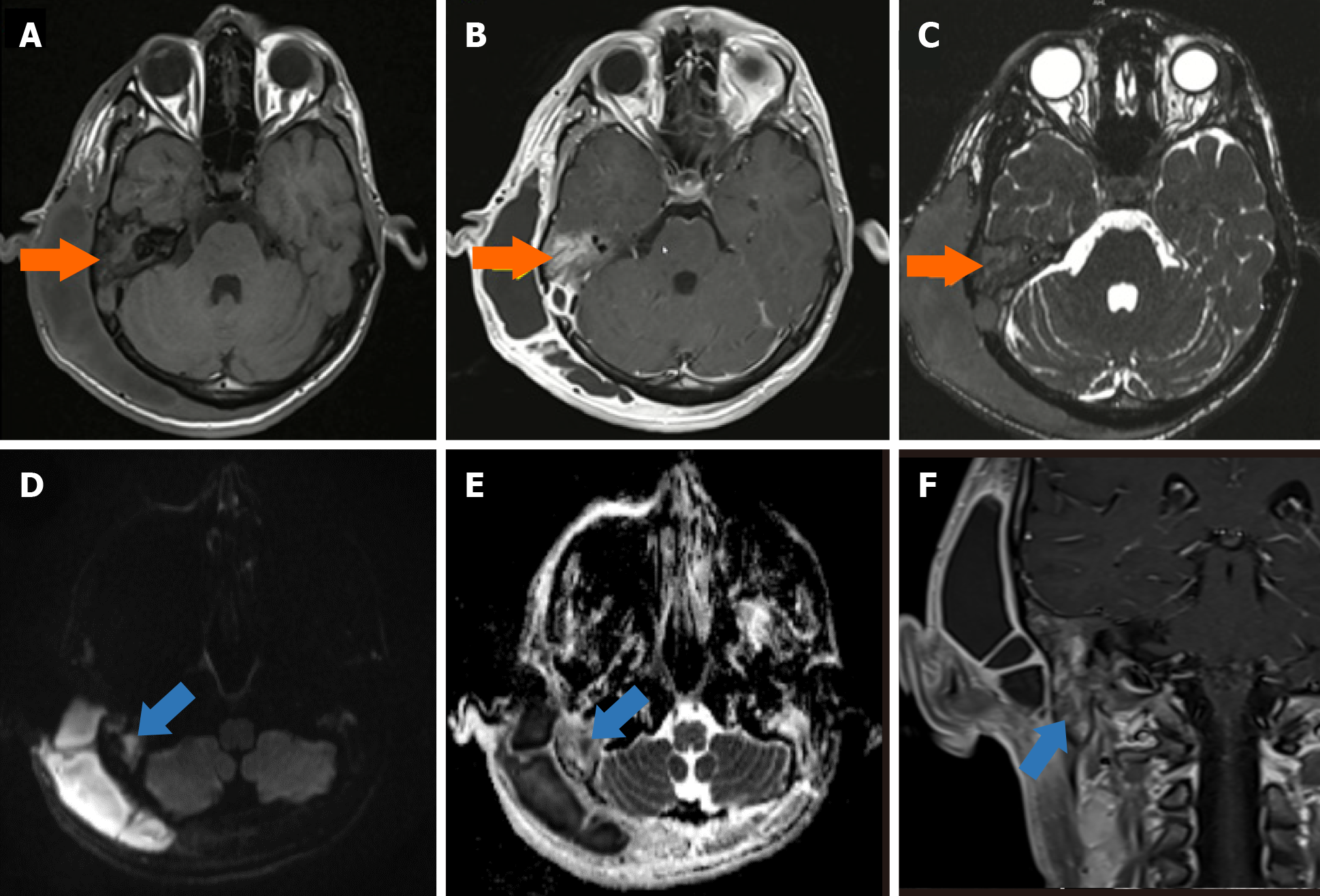
Figure 1 Otomastoiditis and accompanying cholesteatoma.
A: The axial pre-contrast T1W; B: Post-contrast T1W; C: 3D-constructive interference in a steady state MR images show hyper-enhanced inflammatory alterations in the mastoid cells and middle ear on the right side (orange arrows); D: The axial diffusion-weighted; E: Apparent diffusion coefficient map; F: Coronal post-contrast T1W magnetic resonance images show the presence of cholesteatoma in the right inferior mastoid cells, which was not enhanced, but had significant diffusion restriction (blue arrows).
- Citation: Memis KB, Aydin S. Role of imaging in chronic otitis media and its complications. World J Radiol 2025; 17(8): 109447
- URL: https://www.wjgnet.com/1949-8470/full/v17/i8/109447.htm
- DOI: https://dx.doi.org/10.4329/wjr.v17.i8.109447