Copyright
©The Author(s) 2025.
World J Radiol. Jul 28, 2025; 17(7): 107459
Published online Jul 28, 2025. doi: 10.4329/wjr.v17.i7.107459
Published online Jul 28, 2025. doi: 10.4329/wjr.v17.i7.107459
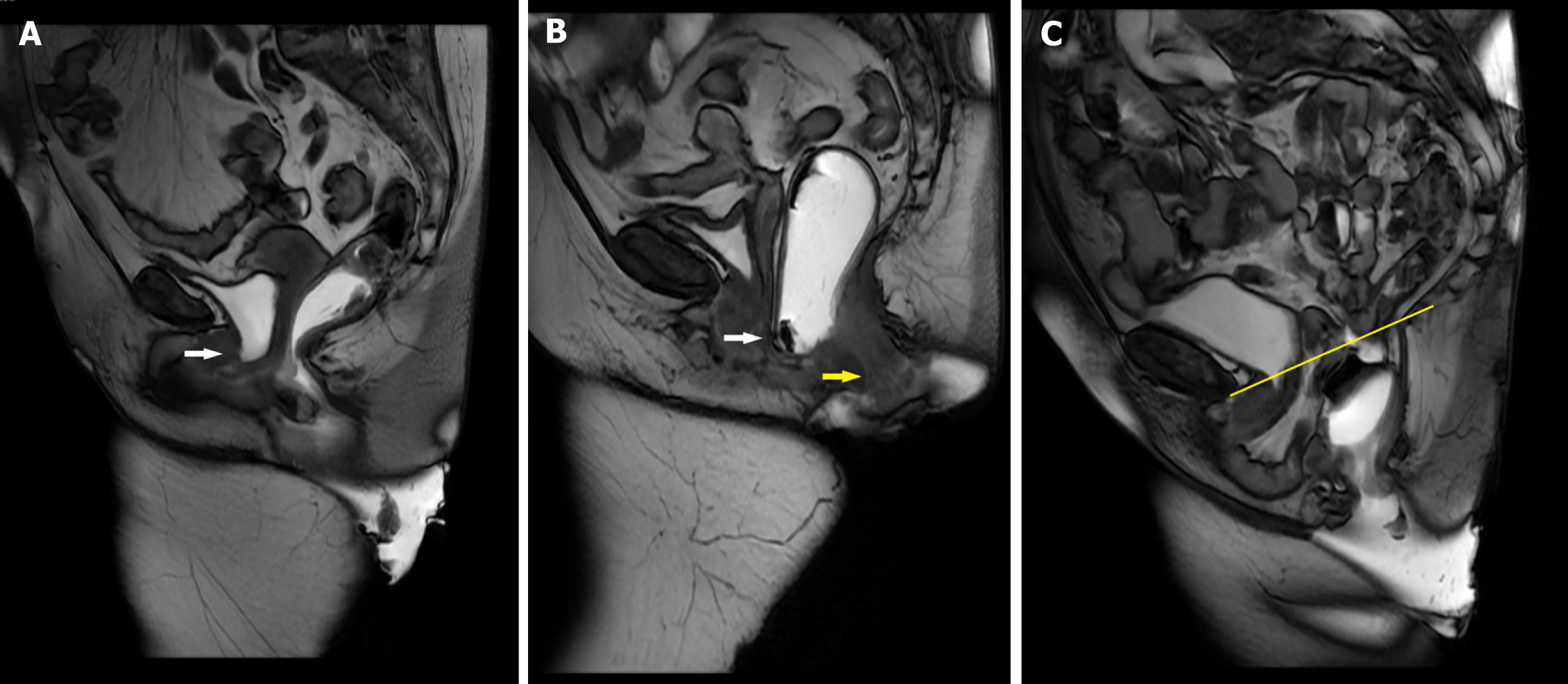
Figure 4 Magnetic resonance defecography images demonstrating.
A: Cystocele inferior aspect of the urinary bladder (arrow) extending caudal to the pubococcygeal line; B: Rectocele/Rectal prolapse-bulging of the anterior rectal wall (white arrow) inferiorly and anteriorly into the posterior vaginal wall. Additionally, there is prolapse of the rectum into the anal canal (yellow arrow) consistent with rectal prolapse; C: Pelvic organ descent-global pelvic floor descent with associated peritoneocele below the pubococcygeal line (yellow line). Image courtesy of Matthew L Osher, MD, Department of Radiology, Henry Ford Providence Hospital.
- Citation: Singh JP, Assaie-Ardakany S, Aleissa MA, Al-Shaer K, Chitragari G, Drelichman ER, Mittal VK, Bhullar JS. Optimizing diagnosis in obstructed defecation syndrome: A review of imaging modalities. World J Radiol 2025; 17(7): 107459
- URL: https://www.wjgnet.com/1949-8470/full/v17/i7/107459.htm
- DOI: https://dx.doi.org/10.4329/wjr.v17.i7.107459


