Copyright
©The Author(s) 2025.
World J Radiol. Jul 28, 2025; 17(7): 107459
Published online Jul 28, 2025. doi: 10.4329/wjr.v17.i7.107459
Published online Jul 28, 2025. doi: 10.4329/wjr.v17.i7.107459
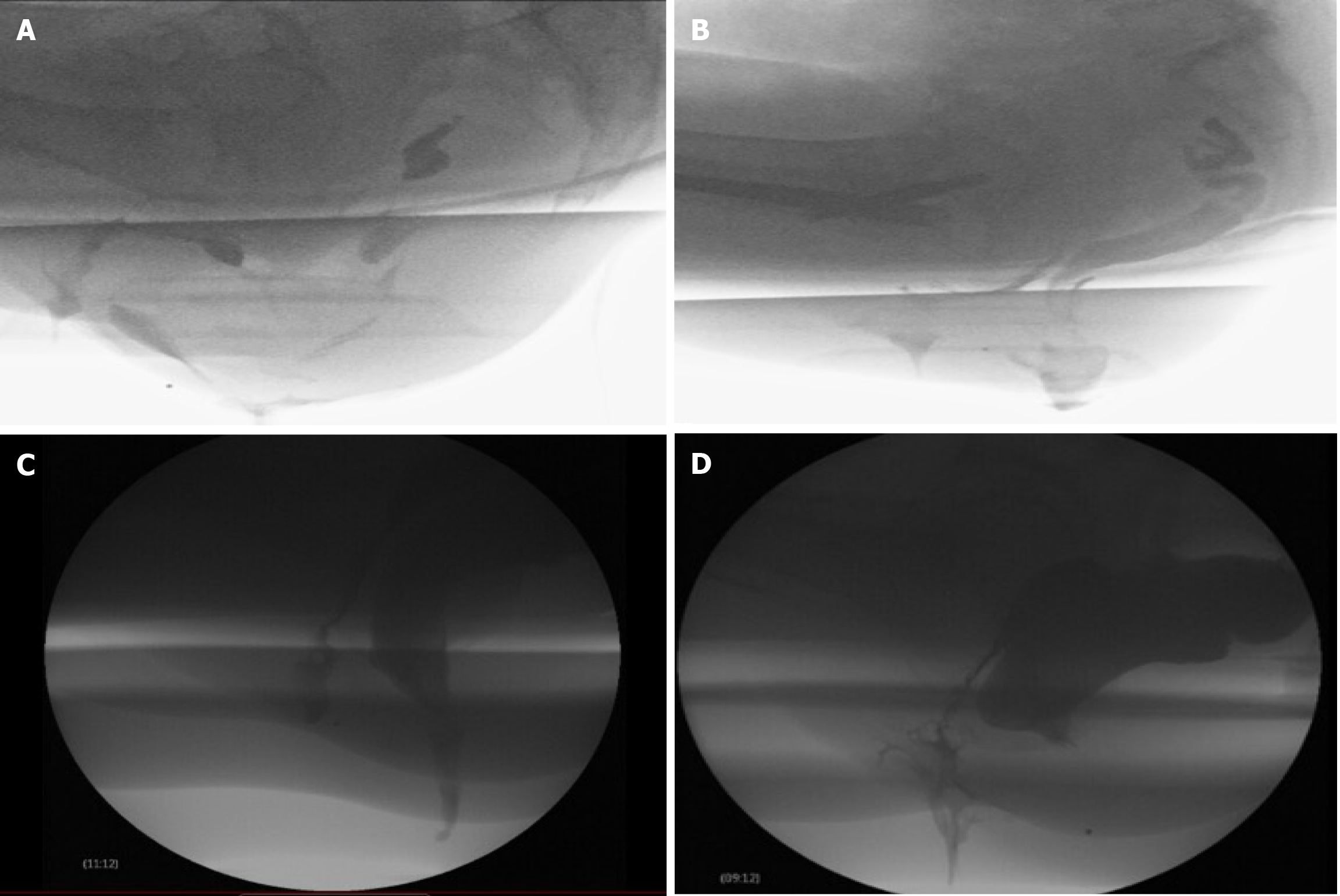
Figure 2 Fluoroscopic defecography images.
A: Rectal intussusception; B: Non-relaxation of the puborectalis muscle, consistent with anismus; C: Fecal incontinence; D: Bulging rectocele into the posterior vaginal wall. Image courtesy of Pamela L Burgess, MD, FACS, Division of Colorectal Surgery, University of Minnesota.
- Citation: Singh JP, Assaie-Ardakany S, Aleissa MA, Al-Shaer K, Chitragari G, Drelichman ER, Mittal VK, Bhullar JS. Optimizing diagnosis in obstructed defecation syndrome: A review of imaging modalities. World J Radiol 2025; 17(7): 107459
- URL: https://www.wjgnet.com/1949-8470/full/v17/i7/107459.htm
- DOI: https://dx.doi.org/10.4329/wjr.v17.i7.107459


