Copyright
©The Author(s) 2025.
World J Radiol. Jul 28, 2025; 17(7): 106556
Published online Jul 28, 2025. doi: 10.4329/wjr.v17.i7.106556
Published online Jul 28, 2025. doi: 10.4329/wjr.v17.i7.106556
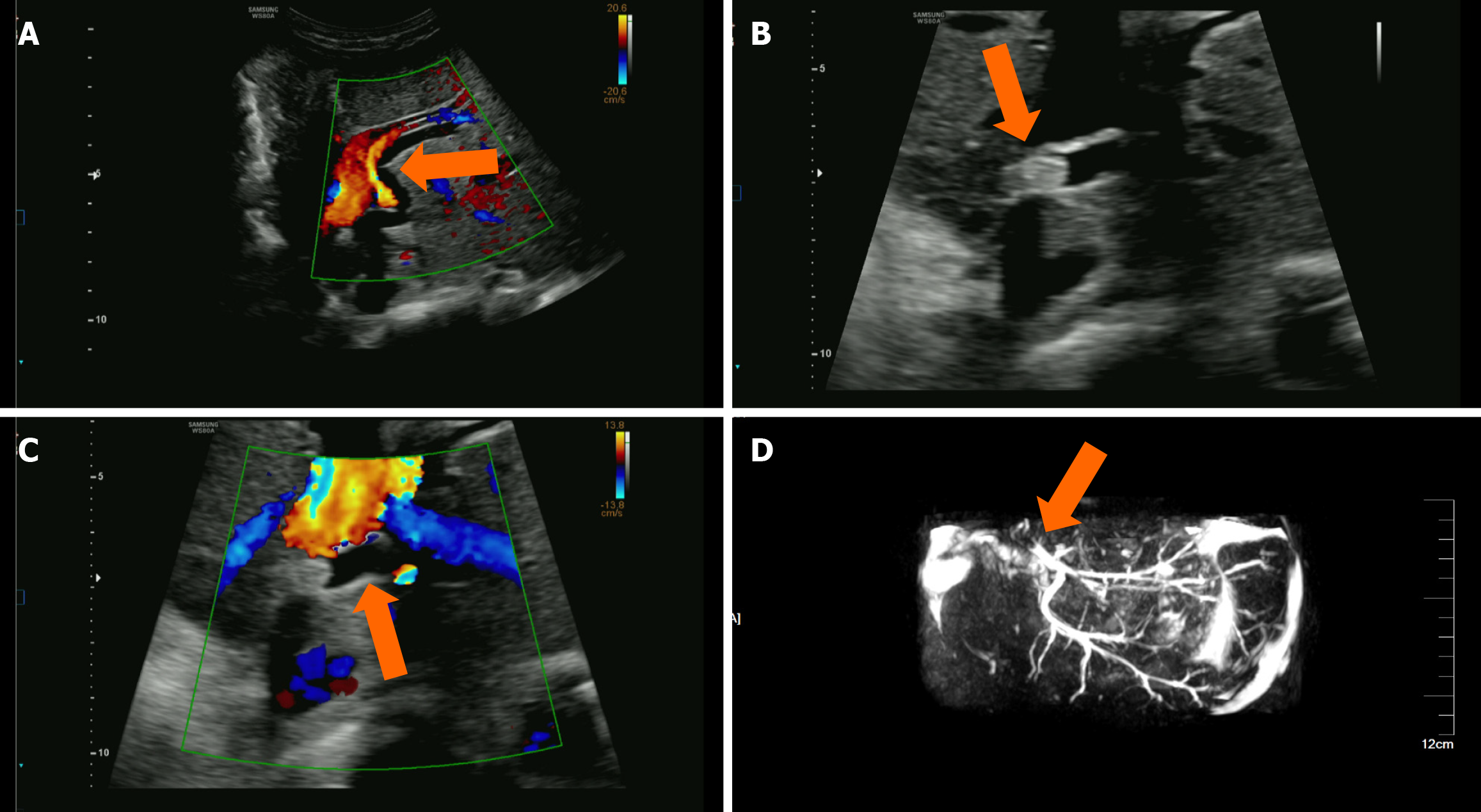
Figure 6 Follow-up ultrasound examination of an 8-year-old girl who underwent split hepatic transplantation due to congenital biliary atresia.
A: Ultrasound showed intrahepatic biliary duct dilation with a smooth biliary duct wall; B and C: Two-dimensional ultrasound showed that a high-echo mass approximately 12 mm × 6 mm in size was visible within the dilated intrahepatic biliary duct, with clear margins from the biliary duct wall and no posterior acoustic shadowing. Color Doppler ultrasound showed no significant color flow signals inside or around the high-echo mass; D: Magnetic resonance cholangiopancreatography showed intrahepatic biliary stones with biliary duct dilation.
- Citation: Zhang Y, Hao J, Luo Z, Li YJ, Liu Z, Zhao NB. Detecting biliary complications following liver transplantation with contrast-enhanced ultrasound. World J Radiol 2025; 17(7): 106556
- URL: https://www.wjgnet.com/1949-8470/full/v17/i7/106556.htm
- DOI: https://dx.doi.org/10.4329/wjr.v17.i7.106556


