Copyright
©The Author(s) 2025.
World J Radiol. Jul 28, 2025; 17(7): 106556
Published online Jul 28, 2025. doi: 10.4329/wjr.v17.i7.106556
Published online Jul 28, 2025. doi: 10.4329/wjr.v17.i7.106556
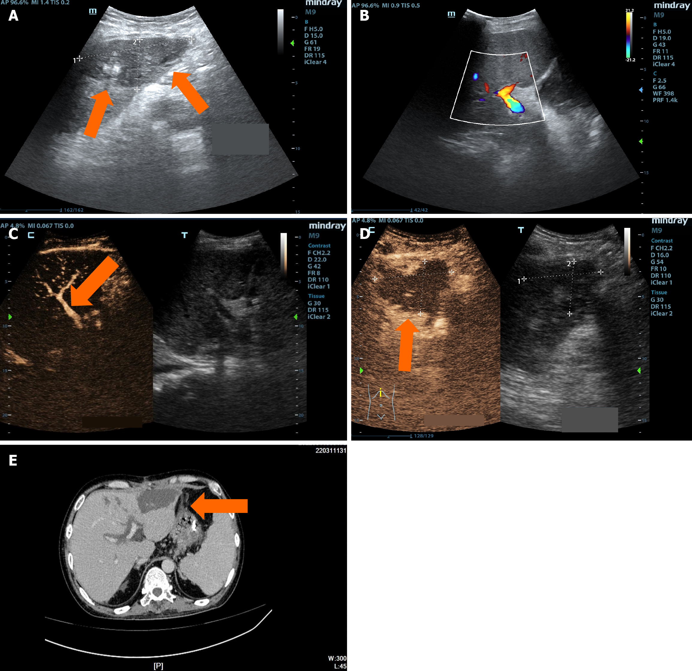
Figure 5 A 30-year-old male patient, more than 4 months post-liver transplantation, presented with a one-week history of fever and diarrhea, followed by vomiting and impaired consciousness for two hours.
A: Two-dimensional ultrasound showed multiple hypoechoic areas within the transplanted liver, suggestive of infarct foci; B: Color Doppler ultrasound showed that no arterial blood flow signals were observed in the hepatic artery course area, indicating hepatic artery occlusion; C: Intravenous contrast-enhanced ultrasound (CEUS) showed no visualization of the entire hepatic artery, consistent with hepatic artery occlusion; D: Intravenous CEUS indicated no enhancement in multiple hypoechoic areas within the liver, consistent with infarct foci; E: Magnetic resonance imaging revealed multiple hypoattenuating lesions within the liver, with an increase in number and size compared to previous scans, suggesting biloma and intrahepatic biliary duct dilation.
- Citation: Zhang Y, Hao J, Luo Z, Li YJ, Liu Z, Zhao NB. Detecting biliary complications following liver transplantation with contrast-enhanced ultrasound. World J Radiol 2025; 17(7): 106556
- URL: https://www.wjgnet.com/1949-8470/full/v17/i7/106556.htm
- DOI: https://dx.doi.org/10.4329/wjr.v17.i7.106556


