Copyright
©The Author(s) 2025.
World J Radiol. Jul 28, 2025; 17(7): 106556
Published online Jul 28, 2025. doi: 10.4329/wjr.v17.i7.106556
Published online Jul 28, 2025. doi: 10.4329/wjr.v17.i7.106556
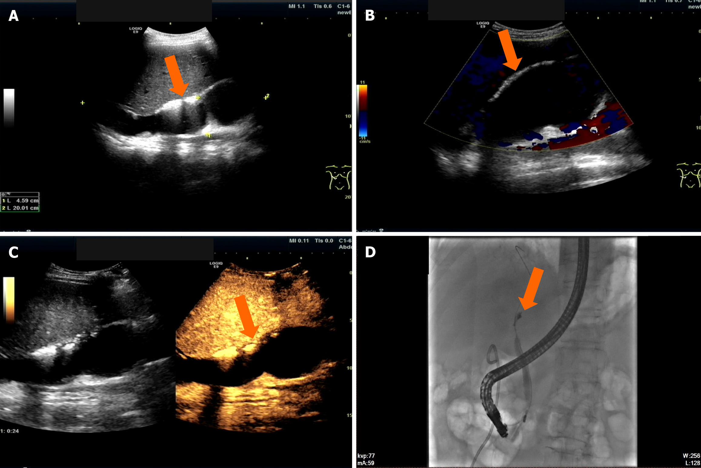
Figure 4 A 60-year-old male patient who underwent liver transplantation for primary biliary cirrhosis.
Twelve days postoperatively, an ultrasound examination revealed a biliary duct fistula. A and B: Two-dimensional ultrasound and color Doppler ultrasound showed that a large anechoic area with an irregular shape and good acoustic transmission was visible in the hepatic hilum, measuring approximately 200 mm × 46 mm, with no significant blood flow signals observed internally; C: Intravenous contrast-enhanced ultrasound showed no internal enhancement, with clear margins (indicated by the arrow); D: Endoscopic retrograde cholangiopancreatography showed that a catheter was successfully inserted, and a small amount of 10% Ultravist (Shering, Berlin, Germany) was injected. A narrow segment, approximately 8 mm long, was visible at the biliary duct anastomosis, and a fistula was observed in the intrahepatic biliary duct on the left side of the liver. The contrast agent infiltrated into the extrahepatic liver through the fistula (indicated by the arrow).
- Citation: Zhang Y, Hao J, Luo Z, Li YJ, Liu Z, Zhao NB. Detecting biliary complications following liver transplantation with contrast-enhanced ultrasound. World J Radiol 2025; 17(7): 106556
- URL: https://www.wjgnet.com/1949-8470/full/v17/i7/106556.htm
- DOI: https://dx.doi.org/10.4329/wjr.v17.i7.106556


