Copyright
©The Author(s) 2025.
World J Radiol. Jun 28, 2025; 17(6): 106682
Published online Jun 28, 2025. doi: 10.4329/wjr.v17.i6.106682
Published online Jun 28, 2025. doi: 10.4329/wjr.v17.i6.106682
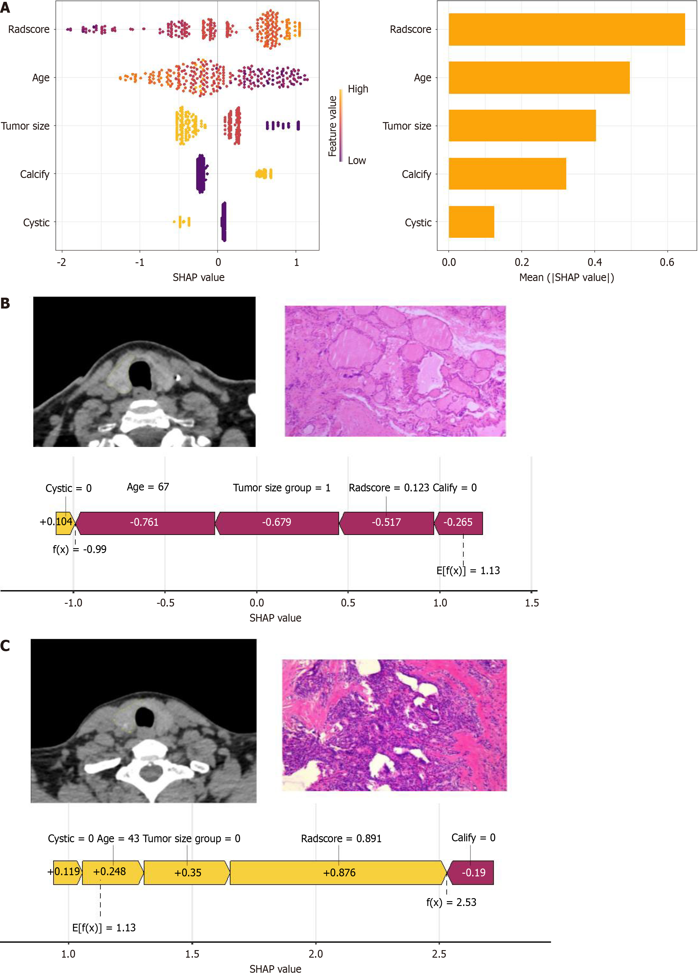
Figure 6 SHapley Additive exPlanations analysis and clinical application.
A: Honeycomb and bar plot ranking SHapley Additive exPlanations values by feature importance; B: Case illustration of a 67-year-old female with an 8 mm right thyroid lobe nodule, pathologically confirmed as nodular goiter; C: Case illustration of a 43-year-old female with a 4 mm right thyroid lobe nodule, pathologically confirmed as papillary thyroid carcinoma. SHAP: SHapley Additive exPlanations.
- Citation: Wang H, Wang X, Du YS, Wang Y, Bai ZJ, Wu D, Tang WL, Zeng HL, Tao J, He J. Non-contrast computed tomography radiomics model to predict benign and malignant thyroid nodules with lobe segmentation: A dual-center study. World J Radiol 2025; 17(6): 106682
- URL: https://www.wjgnet.com/1949-8470/full/v17/i6/106682.htm
- DOI: https://dx.doi.org/10.4329/wjr.v17.i6.106682


