Copyright
©The Author(s) 2025.
World J Radiol. May 28, 2025; 17(5): 106102
Published online May 28, 2025. doi: 10.4329/wjr.v17.i5.106102
Published online May 28, 2025. doi: 10.4329/wjr.v17.i5.106102
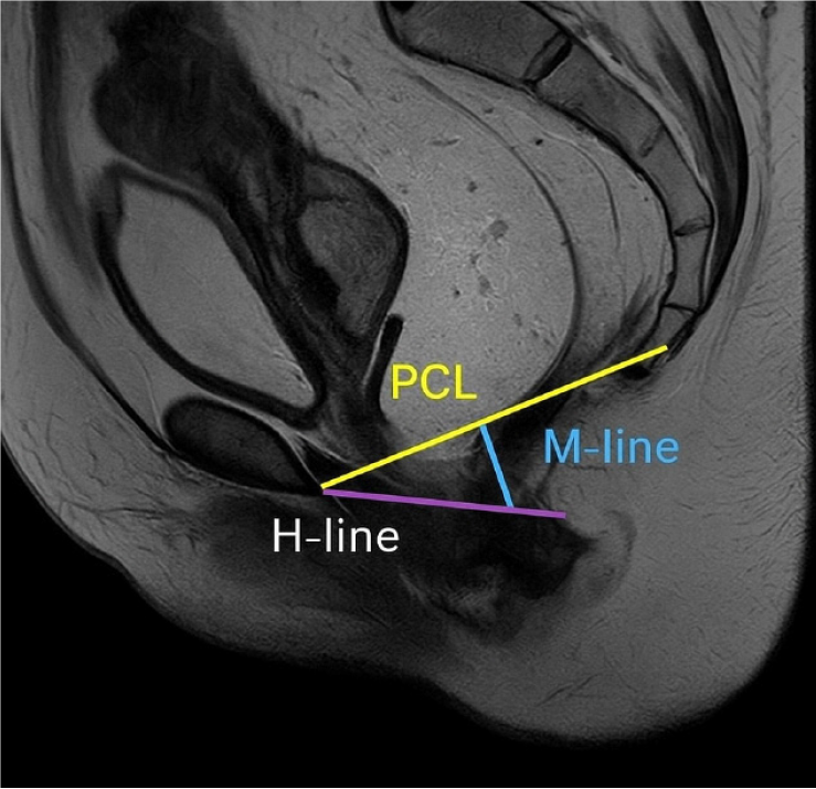
Figure 1 Magnetic resonance defecography T2-weighted image depicting the normal pubococcygeal line[14].
Pubococcygeal line (PCL) (yellow line) spans the inferior border of the pubic symphysis to the last visible coccygeal joint. H-line (purple) represents the length of the anterior-posterior levator hiatus. M-line (blue) measures the vertical descent of the anorectal junction below the PCL. Citation: Korula DR, Chandramohan A, John R, Eapen A. Barium Defecating Proctography and Dynamic Magnetic Resonance Proctography: Their Role and Patient's Perception. J Clin Imaging Sci 2021; 11: 31. Copyright ©The Author(s) 2011. Published by Scientific Scholar LLC (Supplementary material).
- Citation: Or-Rashid MH, Sultana A, Khanduker N, Ony TA, Hossain MM, Rahman J, Chowdhury MZ, Ahmed WU, Uddin MN, Uzzaman MS. Magnetic resonance defecography assessment of obstructed defecation syndrome in patients with chronic constipation in a tertiary care hospital. World J Radiol 2025; 17(5): 106102
- URL: https://www.wjgnet.com/1949-8470/full/v17/i5/106102.htm
- DOI: https://dx.doi.org/10.4329/wjr.v17.i5.106102


