Copyright
©The Author(s) 2025.
World J Radiol. May 28, 2025; 17(5): 106084
Published online May 28, 2025. doi: 10.4329/wjr.v17.i5.106084
Published online May 28, 2025. doi: 10.4329/wjr.v17.i5.106084
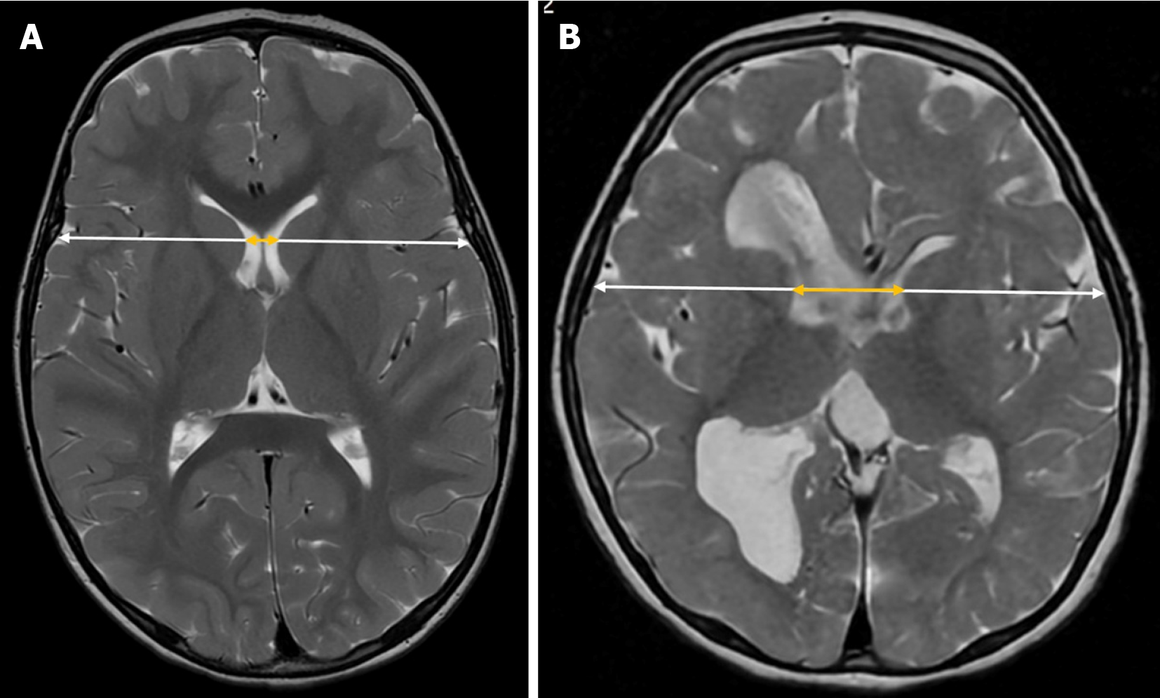
Figure 7 Bicaudate index.
Axial T2-weighted magnetic resonance imaging illustrating the bicaudate index. It is calculated by dividing the distance between the most lateral portions of the frontal horns of the lateral ventricles at the level of the caudate nuclei (yellow arrow) by the transverse brain diameter at the same level (white arrow). A: Normal bicaudate index in a 3-year-old child; B: Increased bicaudate index in a 3-year-old child with asymmetric biventricular hydrocephalus due to perinatal ischemia.
- Citation: Navarro-Ballester A, Álvaro-Ballester R, Lara-Martínez MÁ. Imaging biomarkers for detection and longitudinal monitoring of ventricular abnormalities from birth to childhood. World J Radiol 2025; 17(5): 106084
- URL: https://www.wjgnet.com/1949-8470/full/v17/i5/106084.htm
- DOI: https://dx.doi.org/10.4329/wjr.v17.i5.106084


