Copyright
©The Author(s) 2025.
World J Radiol. May 28, 2025; 17(5): 106084
Published online May 28, 2025. doi: 10.4329/wjr.v17.i5.106084
Published online May 28, 2025. doi: 10.4329/wjr.v17.i5.106084
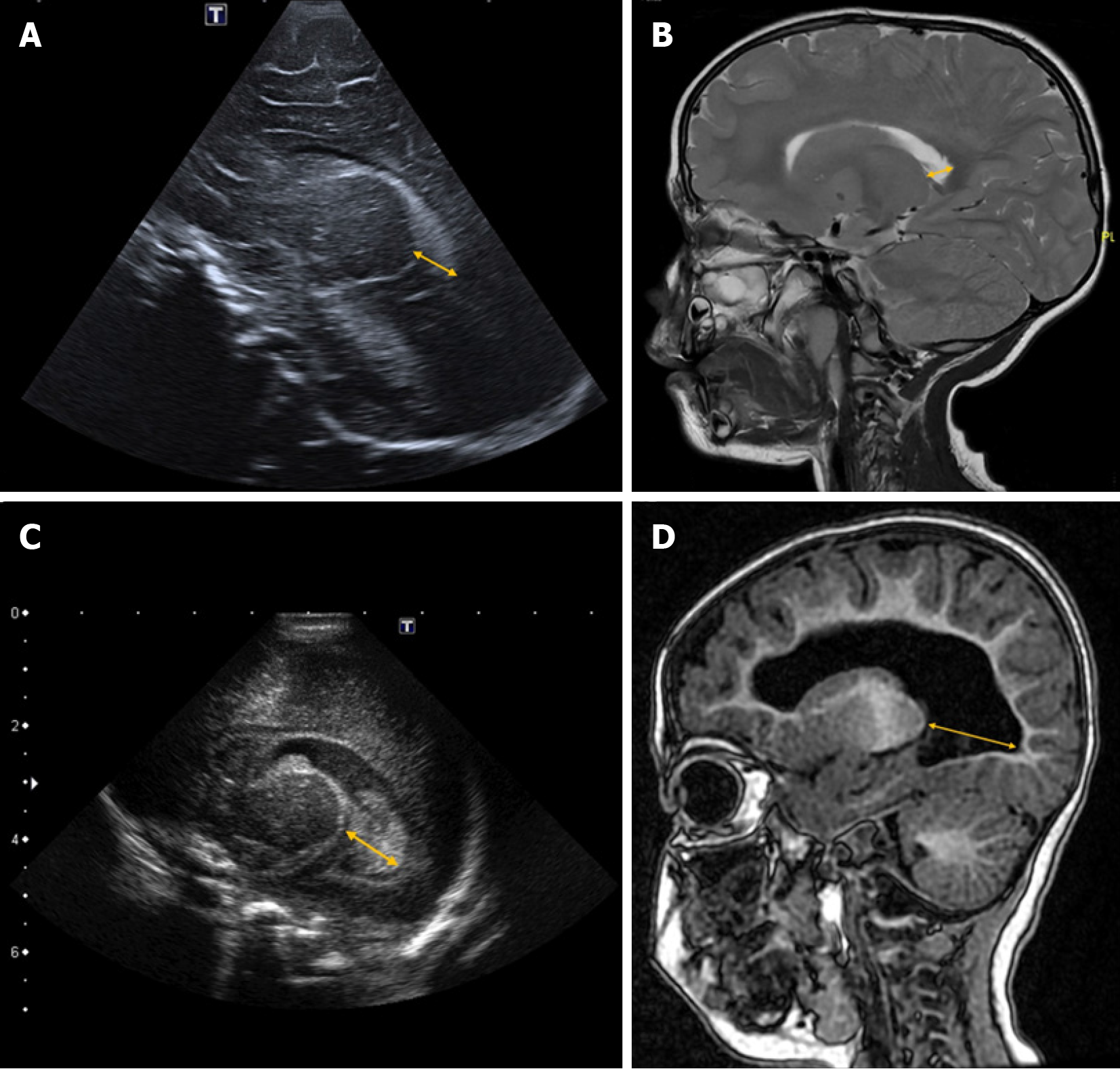
Figure 2 Thalamo-occipital distance.
Parasagittal ultrasound and sagittal magnetic resonance imaging (MRI) images illustrating the thalamo-occipital distance. The measurement corresponds to the distance between the posterior edge of the thalamus at its junction with the choroid plexus and the most external point of the occipital horn of the lateral ventricle (yellow arrow). A: Normal thalamo-occipital distance in a preterm infant (33 + 4 weeks) on parasagittal ultrasound; B: Normal thalamo-occipital distance in a 3-year-old child on sagittal T2-weighted MRI; C: Increased thalamo-occipital distance in a preterm infant (26 + 1 weeks) with hydrocephalus secondary to germinal matrix hemorrhage on parasagittal ultrasound; D: Increased thalamo-occipital distance in a 3-year-old child with asymmetric biventricular hydrocephalus due to perinatal ischemia on sagittal T1-weighted MRI.
- Citation: Navarro-Ballester A, Álvaro-Ballester R, Lara-Martínez MÁ. Imaging biomarkers for detection and longitudinal monitoring of ventricular abnormalities from birth to childhood. World J Radiol 2025; 17(5): 106084
- URL: https://www.wjgnet.com/1949-8470/full/v17/i5/106084.htm
- DOI: https://dx.doi.org/10.4329/wjr.v17.i5.106084


