
















©The Author(s) 2025.
World J Radiol. Dec 28, 2025; 17(12): 114595
Published online Dec 28, 2025. doi: 10.4329/wjr.v17.i12.114595
Published online Dec 28, 2025. doi: 10.4329/wjr.v17.i12.114595
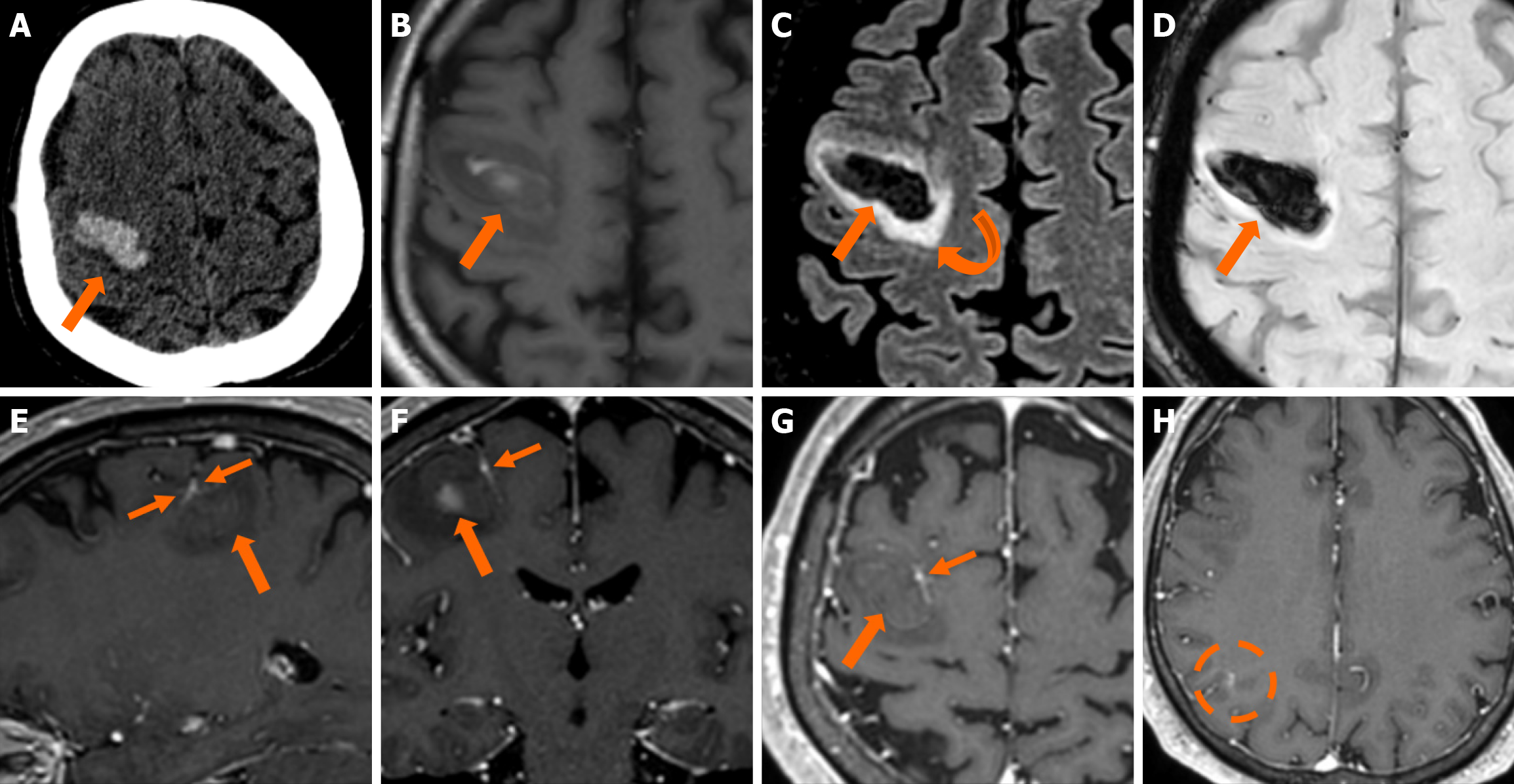
Figure 11 Cerebral cavernous malformation and developmental venous anomaly.
A: Non-contrast-enhanced computed tomography obtained in a 56-year-old man presenting to the emergency department with left-hand weakness shows an intracerebral hematoma in the right frontal lobe (thick arrow). The etiology remained unclear, as no helpful findings were depicted in the computed tomography angiography and venography study performed (not shown); B: On T1-weighted images, the hematoma displays a heterogeneous hyperintense signal due to the recent hemorrhage; C: On fluid-attenuated inversion recovery images, mild surrounding vasogenic edema is also noted (curved arrow); D: Hemosiderin presence is also verified on susceptibility-weighted imaging; E-G: On T1-weighted images sagittal (E), coronal (F) and axial (G) reconstructions following intravenous gadolinium administration, an ectopic vein with “caput medusae” morphology is noted immediately adjacent to the internal border of the hematoma (thin arrows), indicating the presence of a developmental venous anomaly and thus pointing towards an underlying recently bled cerebral cavernous malformation as the underlying etiology for the hematoma; H: An additional developmental venous anomaly was also noted on the same side in a more posterior location (dashed circle).
- Citation: Arkoudis NA, Siderakis M, Tsetsou I, Efthymiou E, Triantafyllou G, Chalmoukis D, Karachaliou A, Papadopoulos A, Prountzos S, Moschovaki-Zeiger O, Gouliopoulos N, Papakonstantinou O, Filippiadis D, Velonakis G. Developmental venous anomalies and cerebral cavernous malformations: Partners in crime. World J Radiol 2025; 17(12): 114595
- URL: https://www.wjgnet.com/1949-8470/full/v17/i12/114595.htm
- DOI: https://dx.doi.org/10.4329/wjr.v17.i12.114595