
















©The Author(s) 2025.
World J Radiol. Dec 28, 2025; 17(12): 114595
Published online Dec 28, 2025. doi: 10.4329/wjr.v17.i12.114595
Published online Dec 28, 2025. doi: 10.4329/wjr.v17.i12.114595
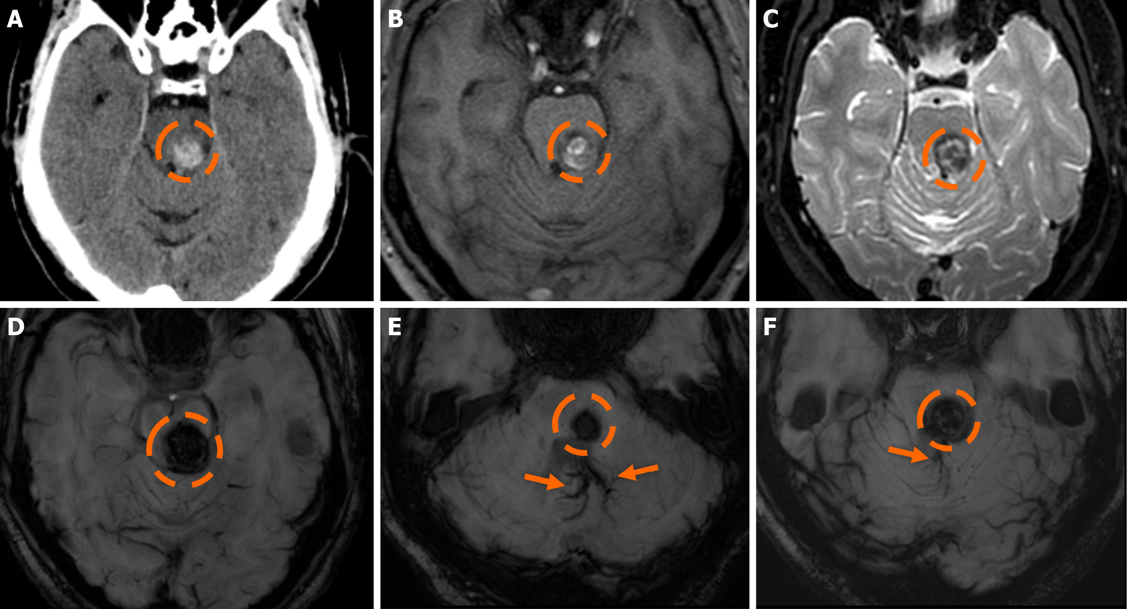
Figure 10 Cerebral cavernous malformation and developmental venous anomaly.
A: Non-contrast-enhanced computed tomography obtained in a 67-year-old man presenting to the emergency department due to impaired balance shows a hyperdense lesion in the pons (dashed circle); B and C: On T1-weighted images the lesion displays a heterogeneous but mostly hyperintense signal, suggesting recent hemorrhage (B), while on T2-weighted images (C), a peripheral low-signal hemosiderin rim is noted, and a “popcorn-like” or “mulberry-like” appearance is depicted centrally; D-F: On susceptibility-weighted imaging, the lesion displayed intense blooming. The findings were mostly suggestive of a cerebral cavernous malformation (Zabramski type I or II, due to mixed findings of a “popcorn” appearance and recent bleed) as the underlying diagnosis, which was further verified by the depiction of a large adjacent developmental venous anomaly (arrows).
- Citation: Arkoudis NA, Siderakis M, Tsetsou I, Efthymiou E, Triantafyllou G, Chalmoukis D, Karachaliou A, Papadopoulos A, Prountzos S, Moschovaki-Zeiger O, Gouliopoulos N, Papakonstantinou O, Filippiadis D, Velonakis G. Developmental venous anomalies and cerebral cavernous malformations: Partners in crime. World J Radiol 2025; 17(12): 114595
- URL: https://www.wjgnet.com/1949-8470/full/v17/i12/114595.htm
- DOI: https://dx.doi.org/10.4329/wjr.v17.i12.114595