
















©The Author(s) 2025.
World J Radiol. Dec 28, 2025; 17(12): 114595
Published online Dec 28, 2025. doi: 10.4329/wjr.v17.i12.114595
Published online Dec 28, 2025. doi: 10.4329/wjr.v17.i12.114595
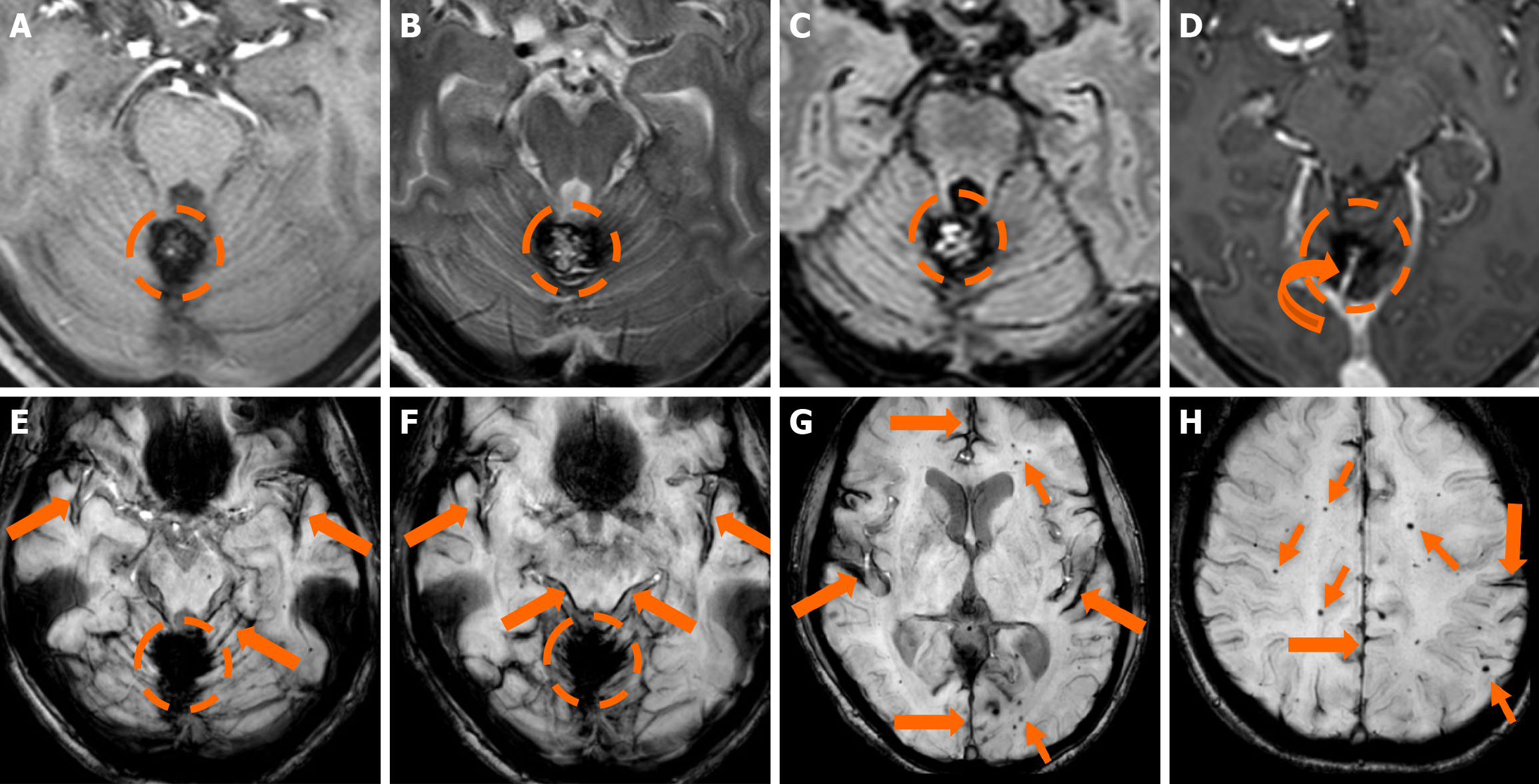
Figure 6 Familial cerebral cavernous malformation syndrome.
Magnetic resonance imaging examination performed due to dizziness in a 50-year-old woman. A-C: A lesion (dashed circle) centered between the cerebellar hemispheres and the scolex is demonstrated. The lesion displays a heterogeneous signal with a few areas of increased signal intensity centrally on T1-weighted images (A), while on T2-weighted (B) and fluid-attenuated inversion recovery (C) images it demonstrates a characteristic “popcorn” appearance with a heterogeneous signal centrally and a low signal hemosiderin rim peripherally; D: T1-weighted axial image following intravenous gadolinium administration demonstrates no discernible enhancement and an ectopic draining vein (curved arrow); E-H: Susceptibility-weighted images verify the hemosiderin presence of the lesion. The findings are suggestive of a Zabramski type II cerebral cavernous malformation with a concomitant developmental venous anomaly (ectopic draining vein) (curved arrow). In addition, susceptibility-weighted images display hemosiderin deposition in the cortical surfaces of the cerebellar and cerebral hemisphere, the pons and midbrain (thick arrows), suggesting superficial siderosis from a prior hemorrhage with subarachnoid extension. Multiple bilateral microbleeds (thin arrows) are also noted, suggesting a diagnosis of familial cerebral cavernous malformation syndrome.
- Citation: Arkoudis NA, Siderakis M, Tsetsou I, Efthymiou E, Triantafyllou G, Chalmoukis D, Karachaliou A, Papadopoulos A, Prountzos S, Moschovaki-Zeiger O, Gouliopoulos N, Papakonstantinou O, Filippiadis D, Velonakis G. Developmental venous anomalies and cerebral cavernous malformations: Partners in crime. World J Radiol 2025; 17(12): 114595
- URL: https://www.wjgnet.com/1949-8470/full/v17/i12/114595.htm
- DOI: https://dx.doi.org/10.4329/wjr.v17.i12.114595