
















©The Author(s) 2025.
World J Radiol. Dec 28, 2025; 17(12): 114595
Published online Dec 28, 2025. doi: 10.4329/wjr.v17.i12.114595
Published online Dec 28, 2025. doi: 10.4329/wjr.v17.i12.114595
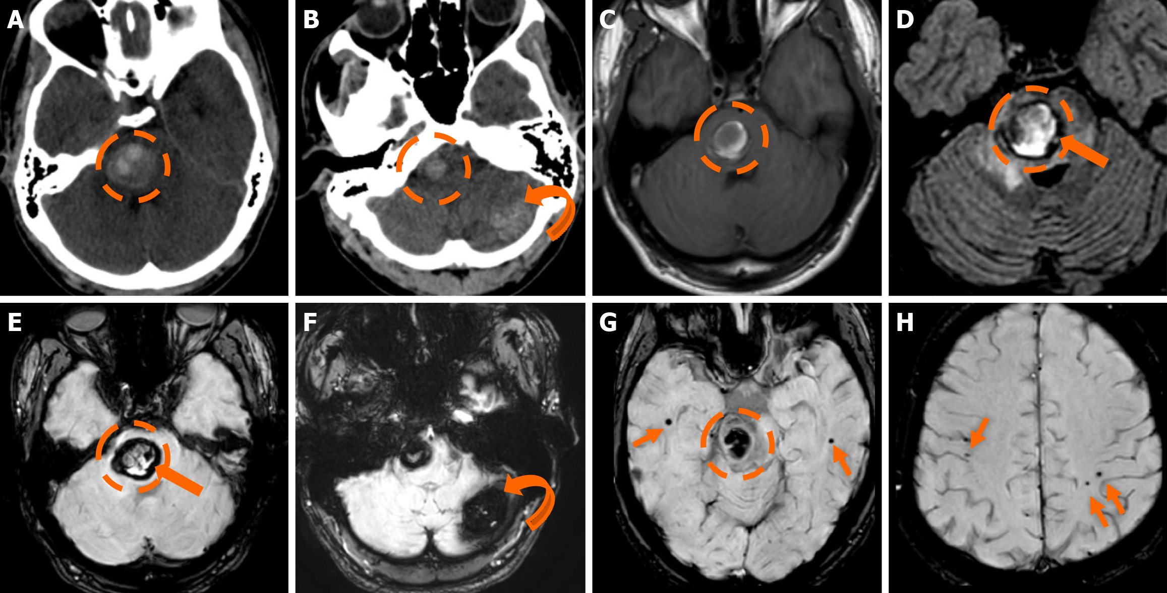
Figure 5 Familial cerebral cavernous malformation syndrome.
A and B: Computed tomography scan performed due to an acute onset headache in a 25-year-old man displayed a heterogeneously hyperdense hemorrhagic lesion in the pons (dashed circle) and an additional mildly hyperdense lesion in the left cerebellar hemisphere (curved arrow); C and D: On the subsequent magnetic resonance imaging examination performed, the pontine lesion (dashed circle) displays a hyperintense signal on T1-weighted (C) and fluid-attenuated inversion recovery images (D) with a low-signal hemosiderin rim (thick arrow); D: Also note the hyperintense signal intensity surrounding the pontine lesion due to vasogenic edema; E-H: Susceptibility-weighted imaging verify the hemosiderin presence and rim (thick arrow) in the pontine lesion (dashed circle) and also display hemosiderin presence with blooming in the left cerebellar hemisphere lesion (curved arrow). Several additional bilateral hemispheric microbleeds are also noted on the susceptibility-weighted images (thin arrows). The findings are suggestive of a Zabramski type I cerebral cavernous malformation (CM) in the pons as indicated by the recent bleed (T1 central hyperintensity) and the surrounding vasogenic edema. The left cerebellar hemispheric lesion represented a Zabramski type II cerebral CM (hemosiderin rim; “popcorn” appearance; blooming; no evidence of recent bleed on the remaining examination). In this setting, the additional bilateral cerebral hemispheric lesions most likely represent Zabramski type IV cerebral CM. The constellation of all the above findings is highly suspicious for familial cerebral CM syndrome.
- Citation: Arkoudis NA, Siderakis M, Tsetsou I, Efthymiou E, Triantafyllou G, Chalmoukis D, Karachaliou A, Papadopoulos A, Prountzos S, Moschovaki-Zeiger O, Gouliopoulos N, Papakonstantinou O, Filippiadis D, Velonakis G. Developmental venous anomalies and cerebral cavernous malformations: Partners in crime. World J Radiol 2025; 17(12): 114595
- URL: https://www.wjgnet.com/1949-8470/full/v17/i12/114595.htm
- DOI: https://dx.doi.org/10.4329/wjr.v17.i12.114595