
















©The Author(s) 2025.
World J Radiol. Dec 28, 2025; 17(12): 114595
Published online Dec 28, 2025. doi: 10.4329/wjr.v17.i12.114595
Published online Dec 28, 2025. doi: 10.4329/wjr.v17.i12.114595
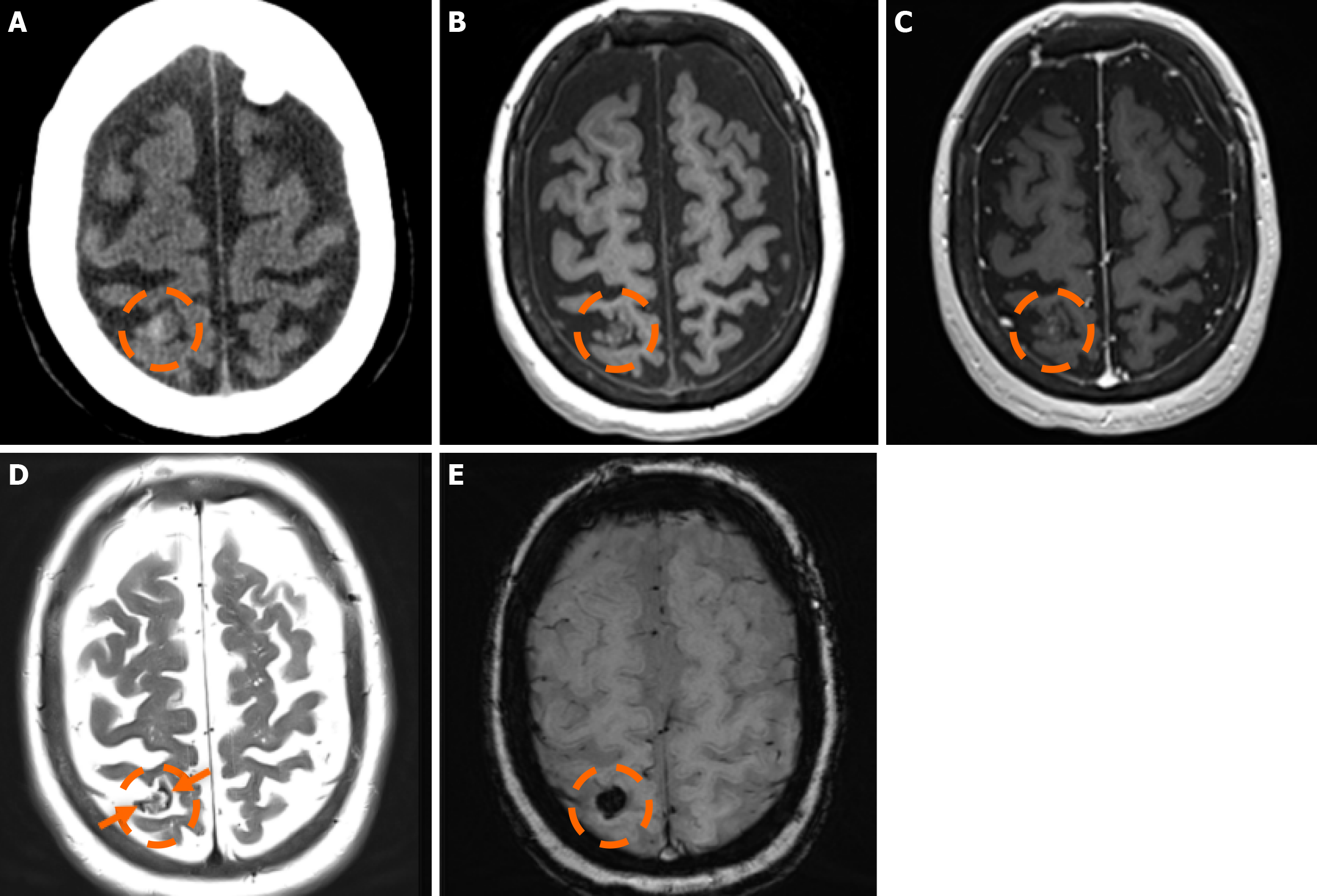
Figure 4 Cerebral cavernous malformation.
A: Computed tomography scan performed in a 65-year-old woman due to memory deficits displayed a mildly hyperdense lesion in the right parietal lobe (dashed circle), which raised suspicion for a hemorrhage; B and C: On the subsequent magnetic resonance imaging examination performed, the lesion (dashed circle) was hypointense on T1-weighted images (B), without significant or discernible contrast enhancement following intravenous gadolinium administration (C); D: On T2-weighted images, the lesion (dashed circle) displays a heterogeneously hyperintense center with a low signal periphery (arrows) compatible with a hemosiderin rim; E: Susceptibility-weighted imaging shows intense blooming of the lesion (dashed circle). The findings were consistent with a cerebral cavernous malformation diagnosis.
- Citation: Arkoudis NA, Siderakis M, Tsetsou I, Efthymiou E, Triantafyllou G, Chalmoukis D, Karachaliou A, Papadopoulos A, Prountzos S, Moschovaki-Zeiger O, Gouliopoulos N, Papakonstantinou O, Filippiadis D, Velonakis G. Developmental venous anomalies and cerebral cavernous malformations: Partners in crime. World J Radiol 2025; 17(12): 114595
- URL: https://www.wjgnet.com/1949-8470/full/v17/i12/114595.htm
- DOI: https://dx.doi.org/10.4329/wjr.v17.i12.114595