











BPG is committed to discovery and dissemination of knowledge
Review
©The Author(s) 2025.
World J Radiol. Dec 28, 2025; 17(12): 114595
Published online Dec 28, 2025. doi: 10.4329/wjr.v17.i12.114595
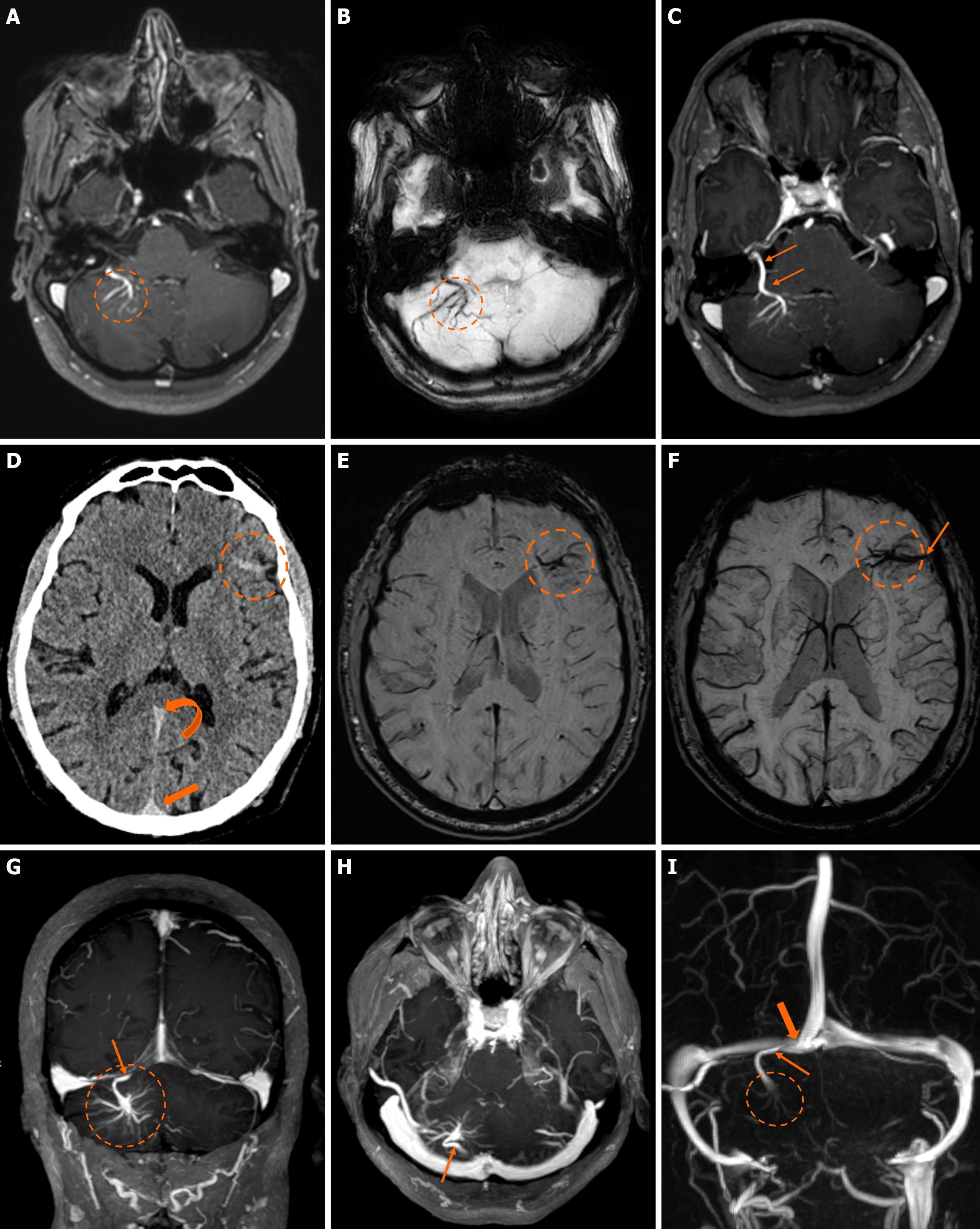
Figure 2
Figure 2 Developmental venous anomalies. A and B: T1-weighted (T1w) axial (A) image following intravenous gadolinium administration, and (B) susceptibility-weighted image demonstrate ectopic veins in the right cerebellar hemisphere (dashed oval); C: Maximum intensity projection of a T1w axial image following intravenous gadolinium administration clearly displays the convergence of the ectopic veins into a singular draining vein (arrow), findings consistent with a developmental venous anomaly; D: Axial non-contrast enhanced computed tomography image performed due to headaches demonstrates hyperdense linear abnormalities in the left frontal lobe (dashed circle) initially perceived as suspicious for subarachnoid hemorrhage. The possibility of the finding representing a vascular structure was also entertained due to the slightly hyperdense appearance of the blood pool in additional vascular structures such as the superior sagittal sinus (thick arrow) and straight sinus (curved arrow); E and F: Susceptibility-weighted axial images from a subsequent magnetic resonance imaging reveal the finding to represent ectopic veins (dashed oval) with a “caput medusae” appearance, converging into a draining vein (thin arrow), findings consistent with a developmental venous anomaly; G-I: Maximum intensity projection of a T1w (G) coronal and axial reconstruction image (H) following intravenous gadolinium administration demonstrate a large developmental venous anomaly in the right cerebellar hemisphere, apparent by the characteristic “caput medusae” (dashed circle) and the draining vein (arrow); I: Maximum intensity projection coronal reconstruction of a phase contrast venography display the collector vein (arrow) draining in the convergence of the right transverse with the superior sagittal dural venous sinus (thick arrow).


Write to the Help Desk