
















©The Author(s) 2025.
World J Radiol. Dec 28, 2025; 17(12): 114595
Published online Dec 28, 2025. doi: 10.4329/wjr.v17.i12.114595
Published online Dec 28, 2025. doi: 10.4329/wjr.v17.i12.114595
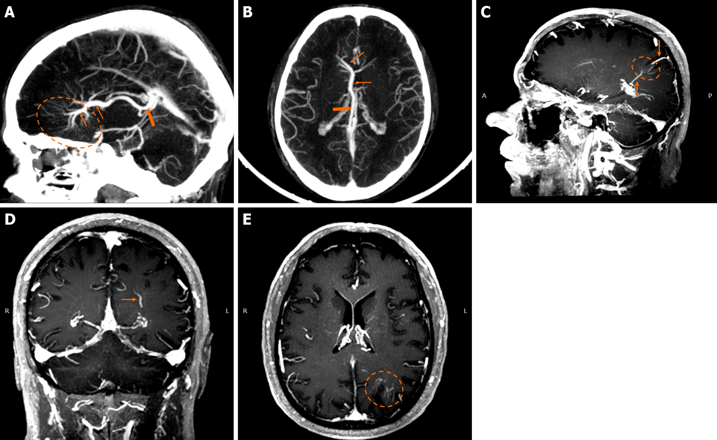
Figure 1 Developmental venous anomalies.
A and B: Sagittal (A) and axial (B) maximum intensity projection reconstruction of a contrast-enhanced computed tomography venography demonstrate a very large developmental venous anomaly in the right frontal lobe, evident by the “caput medusae” morphology (dashed oval) converging in collector veins (thin arrows) and draining in the right internal cerebral vein (thick arrow). Developmental venous anomalies are rarely this large and are usually not easily discernible in computed tomography scans; nonetheless, this example is provided for educational purposes due to its size and characteristic “caput medusae” morphology; C-E: Maximum intensity projection, of T1-weighted sagittal (C), coronal (D) and axial reconstruction images (E) following intravenous gadolinium administration demonstrate a developmental venous anomaly in the left parietal lobe, apparent by the characteristic “caput medusae” (dashed circle) and draining veins (arrows).
- Citation: Arkoudis NA, Siderakis M, Tsetsou I, Efthymiou E, Triantafyllou G, Chalmoukis D, Karachaliou A, Papadopoulos A, Prountzos S, Moschovaki-Zeiger O, Gouliopoulos N, Papakonstantinou O, Filippiadis D, Velonakis G. Developmental venous anomalies and cerebral cavernous malformations: Partners in crime. World J Radiol 2025; 17(12): 114595
- URL: https://www.wjgnet.com/1949-8470/full/v17/i12/114595.htm
- DOI: https://dx.doi.org/10.4329/wjr.v17.i12.114595