
















©The Author(s) 2025.
World J Radiol. Dec 28, 2025; 17(12): 112986
Published online Dec 28, 2025. doi: 10.4329/wjr.v17.i12.112986
Published online Dec 28, 2025. doi: 10.4329/wjr.v17.i12.112986
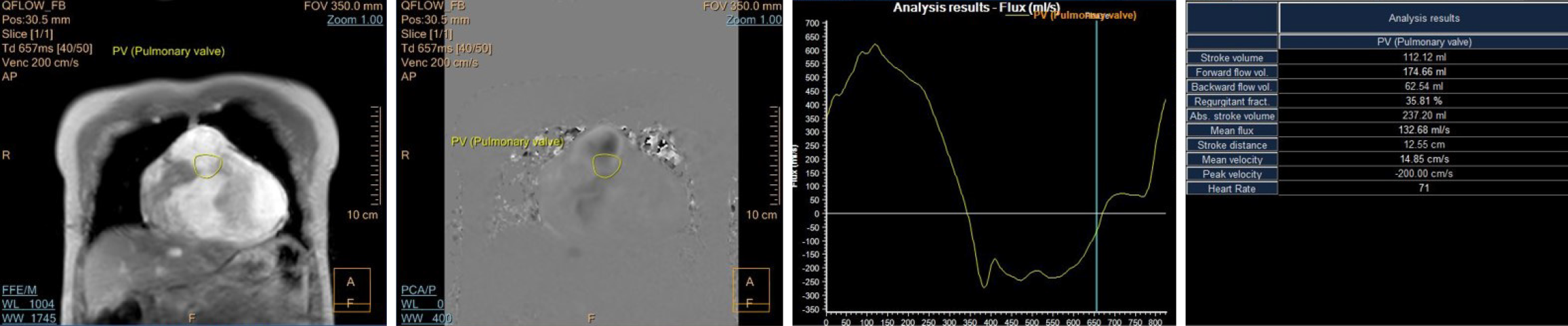
Figure 3 Cardiac magnetic resonance imaging with phase-contrast imaging of the pulmonary valve (region of interest represented by a yellow circle) showing the flow curve with antegrade flow above the baseline and retrograde flow below the baseline, representing the pulmonary regurgitation.
The absolute retrograde flow volume was 62.54 mL, and the regurgitant fraction was calculated by the software as 35.81%.
- Citation: Sharma A, Subramanian P, Soundararajan R, Maralakunte M, Mallick A, Debi U, Jaswal V, Manoj Kumar R, Singhal M. Imaging considerations in tetralogy of Fallot: A comprehensive review. World J Radiol 2025; 17(12): 112986
- URL: https://www.wjgnet.com/1949-8470/full/v17/i12/112986.htm
- DOI: https://dx.doi.org/10.4329/wjr.v17.i12.112986