
















©The Author(s) 2025.
World J Radiol. Dec 28, 2025; 17(12): 112986
Published online Dec 28, 2025. doi: 10.4329/wjr.v17.i12.112986
Published online Dec 28, 2025. doi: 10.4329/wjr.v17.i12.112986
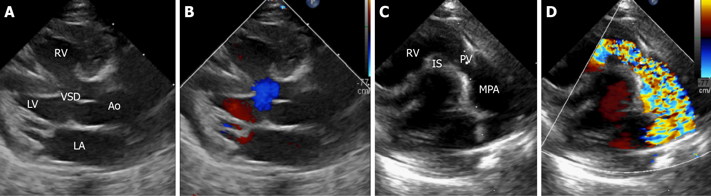
Figure 2 Trans-thoracic echocardiography in classical tetralogy of Fallot.
A and B: Parasternal long axis view (grayscale and Doppler images, respectively) focusing on the aortic valve showing the large ventricular septal defect, aortic override, and right-to-left shunt, blue color in (B); C and D: Parasternal long axis view (grayscale and Doppler images, respectively), focusing on the pulmonary valve showing the turbulence, mosaic pattern in (D) in blood flow due to right ventricular outflow tract obstruction. Ao: Aorta; VSD: Ventricular septal defect; RV: Right ventricle; LV: Left ventricle; MPA: Main pulmonary artery; PV: Pulmonary valve; IS: Infundibular septum.
- Citation: Sharma A, Subramanian P, Soundararajan R, Maralakunte M, Mallick A, Debi U, Jaswal V, Manoj Kumar R, Singhal M. Imaging considerations in tetralogy of Fallot: A comprehensive review. World J Radiol 2025; 17(12): 112986
- URL: https://www.wjgnet.com/1949-8470/full/v17/i12/112986.htm
- DOI: https://dx.doi.org/10.4329/wjr.v17.i12.112986