
















©The Author(s) 2025.
World J Radiol. Dec 28, 2025; 17(12): 112911
Published online Dec 28, 2025. doi: 10.4329/wjr.v17.i12.112911
Published online Dec 28, 2025. doi: 10.4329/wjr.v17.i12.112911
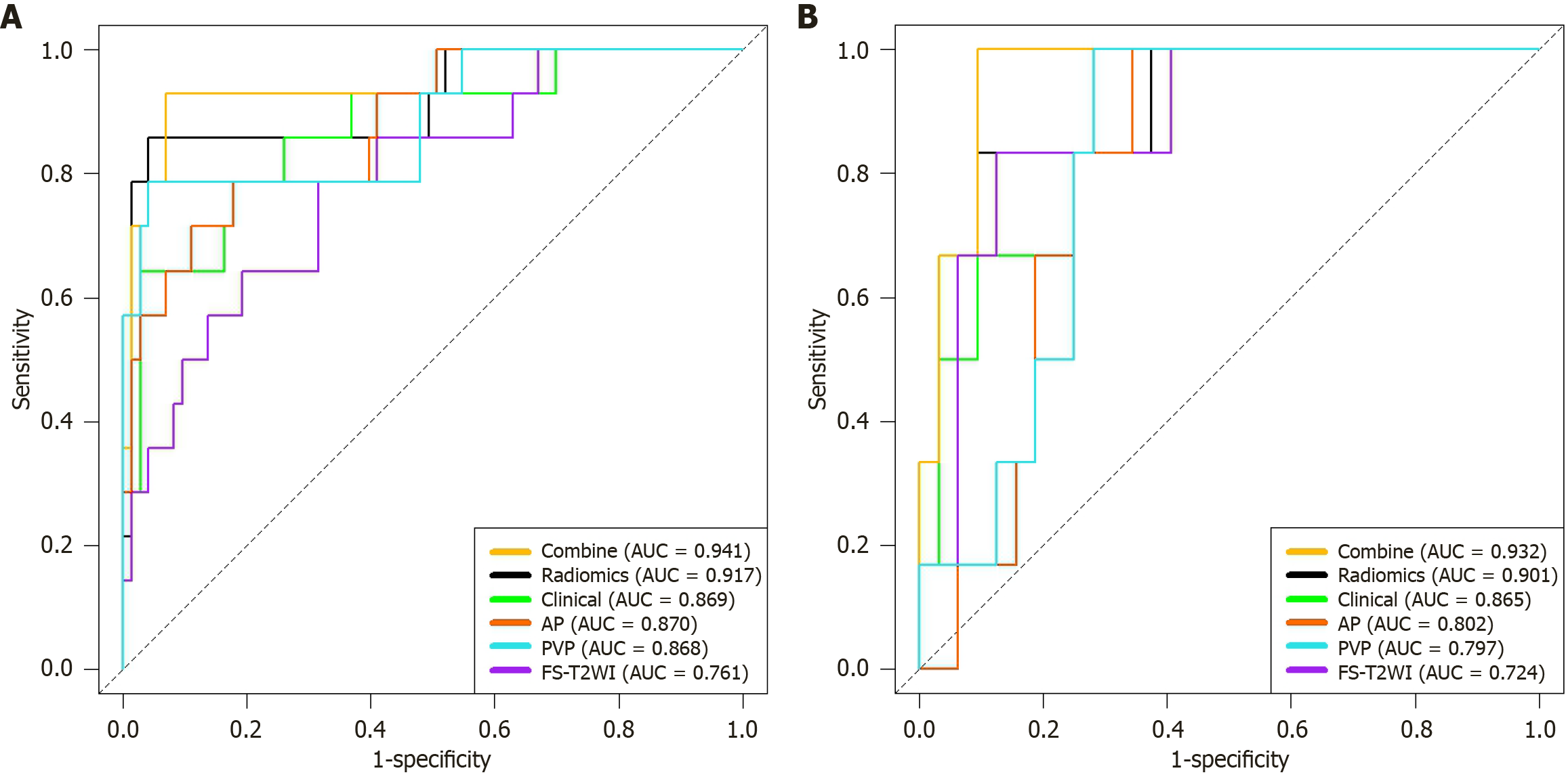
Figure 5 Receiver operating characteristic curves for each model.
A: Training group; B: Validation group. AUC: Area under the receiver operating characteristic curve; AP: Arterial phase; PVP: Portal venous phase; FS-T2WI: Fat-suppressed T2-weighted imaging.
- Citation: Shi Y, Zhang P, Li L, Yang HM, Li ZM, Zheng J, Yang L. Interpretable model based on multisequence magnetic resonance imaging radiomics for predicting the pathological grades of hepatocellular carcinomas. World J Radiol 2025; 17(12): 112911
- URL: https://www.wjgnet.com/1949-8470/full/v17/i12/112911.htm
- DOI: https://dx.doi.org/10.4329/wjr.v17.i12.112911