
















©The Author(s) 2025.
World J Radiol. Nov 28, 2025; 17(11): 114754
Published online Nov 28, 2025. doi: 10.4329/wjr.v17.i11.114754
Published online Nov 28, 2025. doi: 10.4329/wjr.v17.i11.114754
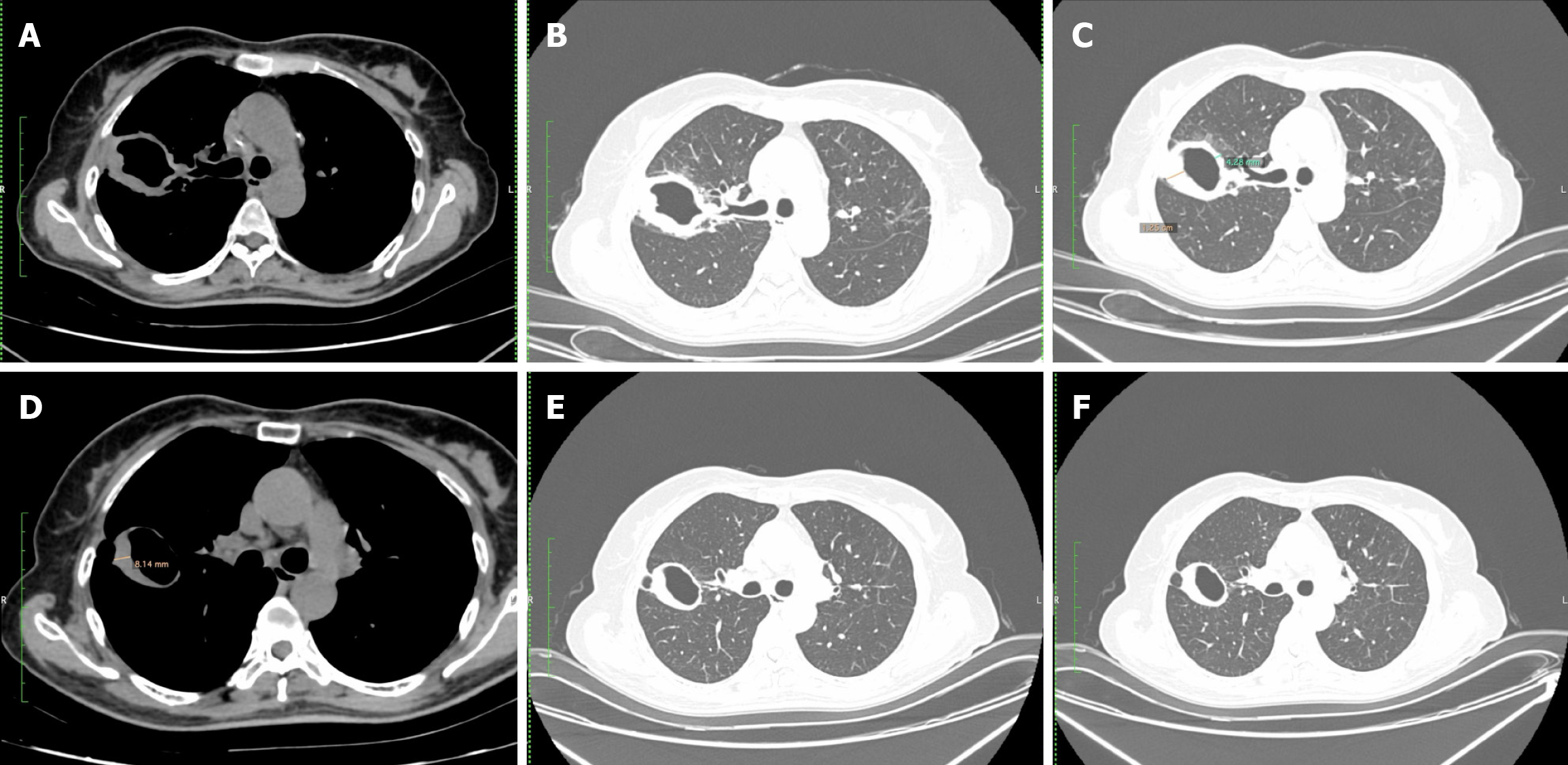
Figure 8 Axial computed tomography images in soft tissue window and lung window in a 59-year-old female, with sputum culture positive for mycobacterium tuberculosis.
A-C: Demonstrates a thick-walled cavity in the right upper lobe. After 4 months of anti-tuberculosis treatment; D-F: Reduced wall thickness of the right upper lobe cavity is noted (from 12.5 mm to 8.14 mm) and the cavity appears much smoother. (Image courtesy Dr. Anagha Joshi, Prof and HOD, Radiology, LTMMC and LTMGH, Mumbai 400022).
- Citation: Merchant SA, Merchant N, Varghese SL, Shaikh MJS. Large language models and large concept models in radiology: Present challenges, future directions, and critical perspectives. World J Radiol 2025; 17(11): 114754
- URL: https://www.wjgnet.com/1949-8470/full/v17/i11/114754.htm
- DOI: https://dx.doi.org/10.4329/wjr.v17.i11.114754