
















©The Author(s) 2025.
World J Radiol. Nov 28, 2025; 17(11): 114754
Published online Nov 28, 2025. doi: 10.4329/wjr.v17.i11.114754
Published online Nov 28, 2025. doi: 10.4329/wjr.v17.i11.114754
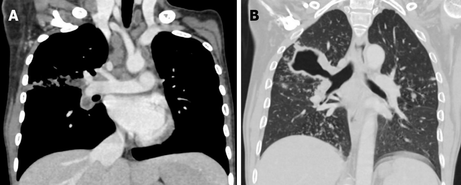
Figure 7 Computed tomography image.
A: Childhood tuberculosis primary Ghons’ complex computed tomography appearance: Laterally positioned Ghon foci with sub-pleural involvement, with their draining lymphatics heading all the way up to right hilar lymphadenopathy. Additionally note mediastinal lymphadenopathy (many lymph nodes reveal necrotic areas); B: Computed tomography image: Large thick-walled tuberculosis cavity communicating with the right main bronchus in a multidrug-resistant patient. (Image courtesy Dr. Anagha Joshi, Prof and HOD, Radiology, LTMMC and LTMGH, Mumbai 400022).
- Citation: Merchant SA, Merchant N, Varghese SL, Shaikh MJS. Large language models and large concept models in radiology: Present challenges, future directions, and critical perspectives. World J Radiol 2025; 17(11): 114754
- URL: https://www.wjgnet.com/1949-8470/full/v17/i11/114754.htm
- DOI: https://dx.doi.org/10.4329/wjr.v17.i11.114754