
















©The Author(s) 2025.
World J Radiol. Nov 28, 2025; 17(11): 114754
Published online Nov 28, 2025. doi: 10.4329/wjr.v17.i11.114754
Published online Nov 28, 2025. doi: 10.4329/wjr.v17.i11.114754
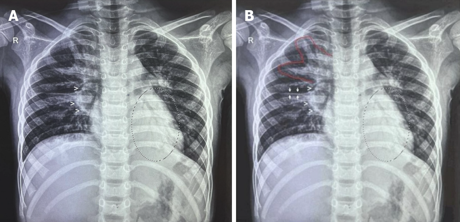
Figure 6 Tuberculosis primary complex - the classic Ghon complex.
A: As seen in in a 5-year-old child: (1) Multiple: Ghon foci in the right upper lobe; (2) Draining lymphatics heading towards; (3) Right hilar lymphadenopathy (white open arrowheads); and (4) Minimal (lamellar) effusion in right horizontal fissure with minimal blunting of the right costo-phrenic angle. Additionally, a retrocardiac air bronchogram indicates tuberculosis (TB) broncho pneumonia (circled area) and hepatomegaly are noted. (Image courtesy Dr. Prakash V Vaidya, Child Health Clinic, Mumbai and Sr. Consultant Pediatrician, Fortis Hospital, Mumbai 400080); B: TB primary complex in a 5-year-old child: The classic Ghon Complex: (1) Multiple: Ghon foci in the rt. upper lobe; (2) Draining lymphatics [(1) and (2) are the areas marked with a red outline]; (3) Heading towards right. hilar lymphadenopathy (large white open arrowheads); and (4) Minimal (lamellar) effusion in right horizontal fissure (white arrows) with minimal blunting of the right costo-phrenic angle. Additionally, a retrocardiac air bronchogram indicates TB broncho pneumonia (circled area), and hepatomegaly are noted. (Image courtesy Dr. Prakash V Vaidya, Child Health Clinic, Mumbai and Sr. Consultant Pediatrician, Fortis Hospital, Mumbai 400080).
- Citation: Merchant SA, Merchant N, Varghese SL, Shaikh MJS. Large language models and large concept models in radiology: Present challenges, future directions, and critical perspectives. World J Radiol 2025; 17(11): 114754
- URL: https://www.wjgnet.com/1949-8470/full/v17/i11/114754.htm
- DOI: https://dx.doi.org/10.4329/wjr.v17.i11.114754