
















©The Author(s) 2025.
World J Radiol. Nov 28, 2025; 17(11): 113701
Published online Nov 28, 2025. doi: 10.4329/wjr.v17.i11.113701
Published online Nov 28, 2025. doi: 10.4329/wjr.v17.i11.113701
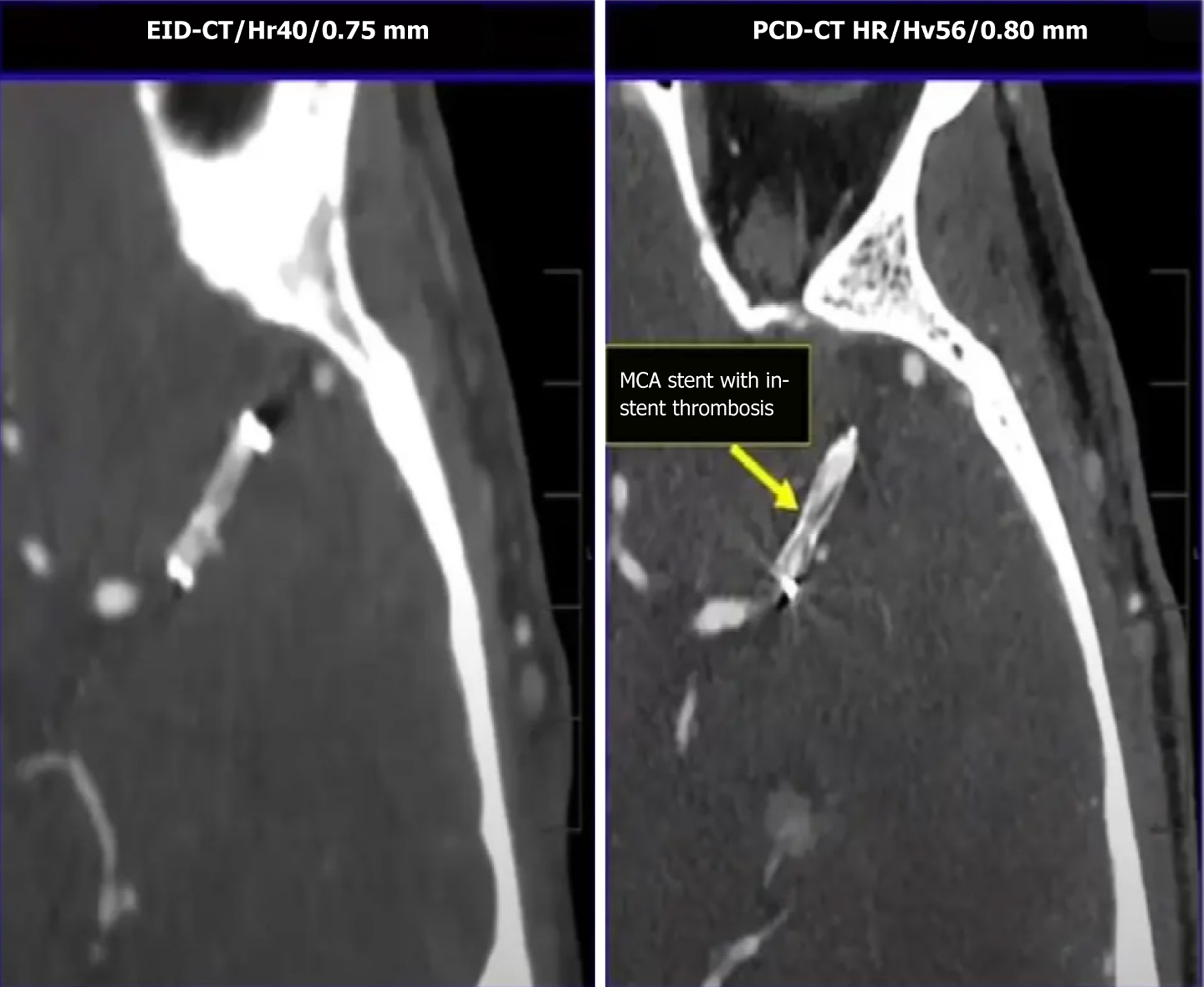
Figure 3 Image showing a conventional image of a head computed tomography angiogram (on the left) and its corresponding photon-counting computed tomography version (on the right), which clearly shows a middle cerebral artery stent with in-stent thrombosis.
EID-CT: Energy-integrating detector-computed tomography; PCD-CT: Photon-counting detector-computed tomography; MCA: Middle cerebral artery. Image courtesy of Professor Cynthia H McCollough, Mayo Clinic, Rochester, MN, United States.
- Citation: Perera Molligoda Arachchige AS, Alves GAM, Fedorov D, Ressa G, Cappellini L, Levi R, Savini G, Catapano F, Francone M, Politi LS. Neuroimaging with photon-counting computed tomography: A review of clinical applications. World J Radiol 2025; 17(11): 113701
- URL: https://www.wjgnet.com/1949-8470/full/v17/i11/113701.htm
- DOI: https://dx.doi.org/10.4329/wjr.v17.i11.113701