
















©The Author(s) 2025.
World J Radiol. Nov 28, 2025; 17(11): 113153
Published online Nov 28, 2025. doi: 10.4329/wjr.v17.i11.113153
Published online Nov 28, 2025. doi: 10.4329/wjr.v17.i11.113153
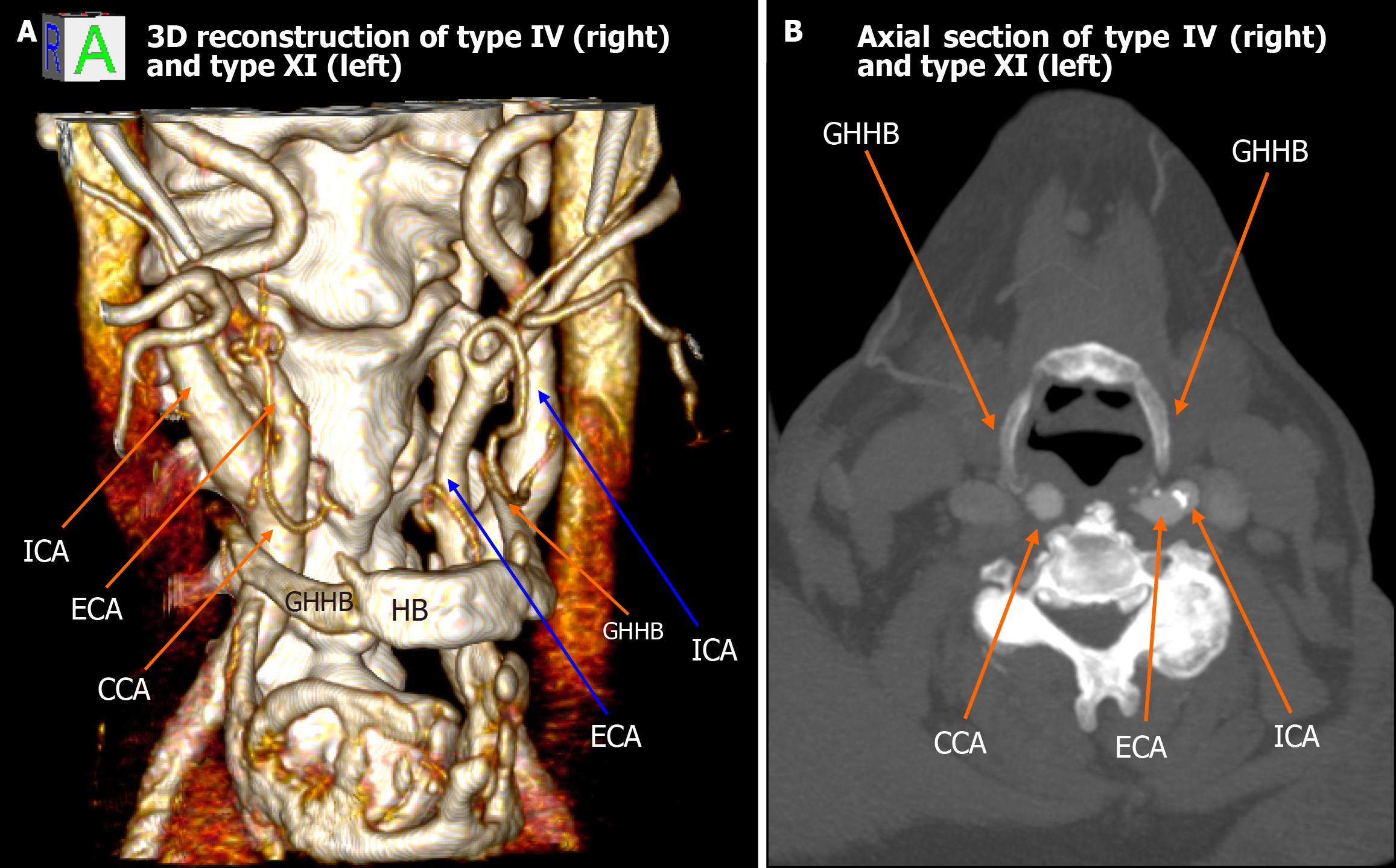
Figure 2 Type IV (right) and type XI (left) configuration.
A: 3D reconstruction shows the right common carotid artery positioned medial to the greater horn of hyoid bone (type IV) and the left external carotid artery lateral with internal carotid artery medial to greater horn of hyoid bone (type XI); B: Axial computed tomography images confirm the asymmetric carotid-hyoid relationships. ICA: Internal carotid artery; ECA: External carotid artery; CCA: Common carotid artery; GHHB: Greater horn of hyoid bone; HB: Hyoid bone.
- Citation: Karangeli N, Triantafyllou G, Papadopoulos-Manolarakis P, Arkoudis NA, Velonakis G, Samolis A, Piagkou M. Variations in the spatial relationship between the hyoid bone and the carotid arteries and their clinical significance. World J Radiol 2025; 17(11): 113153
- URL: https://www.wjgnet.com/1949-8470/full/v17/i11/113153.htm
- DOI: https://dx.doi.org/10.4329/wjr.v17.i11.113153