
















©The Author(s) 2025.
World J Radiol. Nov 28, 2025; 17(11): 113153
Published online Nov 28, 2025. doi: 10.4329/wjr.v17.i11.113153
Published online Nov 28, 2025. doi: 10.4329/wjr.v17.i11.113153
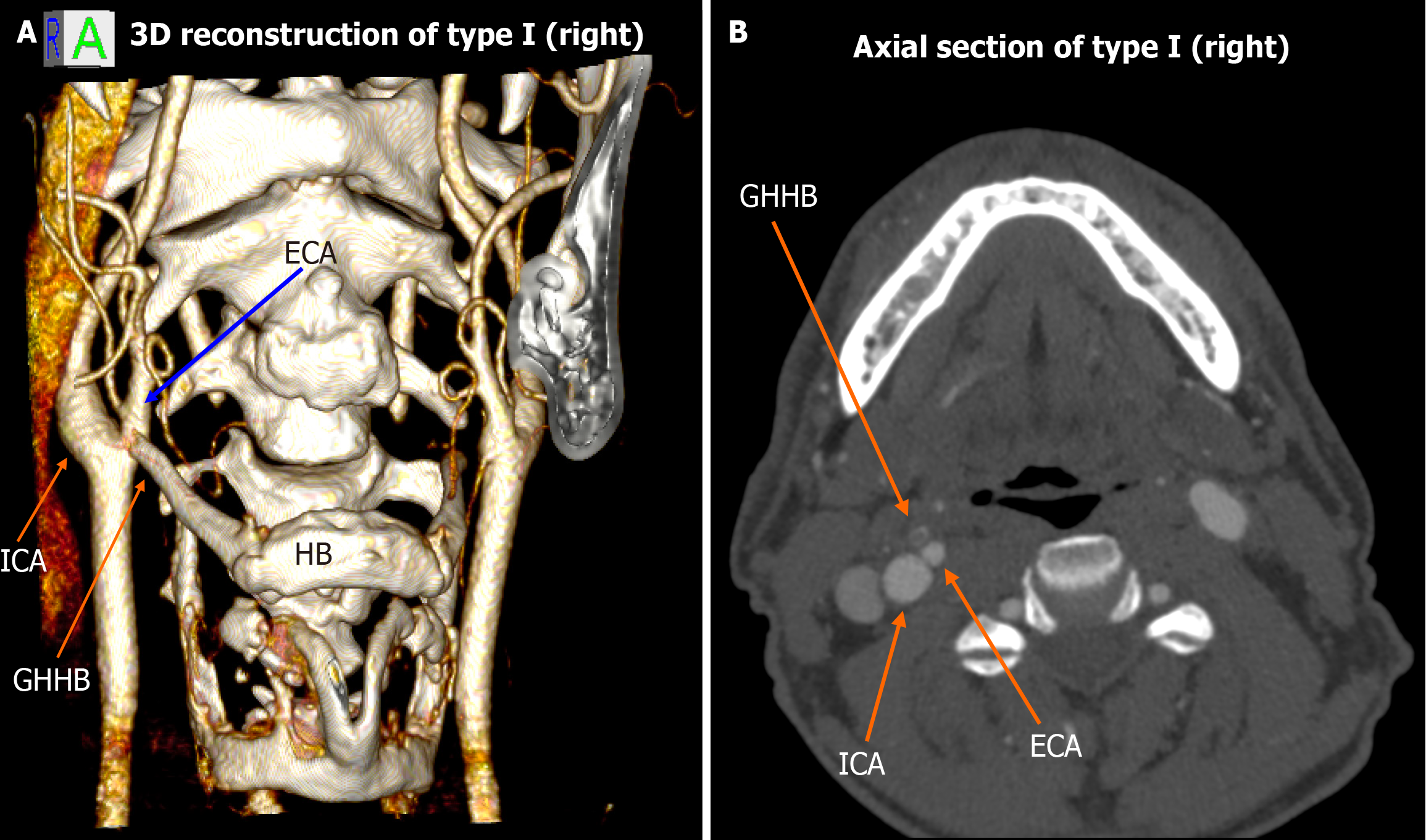
Figure 1 Type I spatial configuration (right-sided).
A: 3D volume rendering shows the external carotid artery medial to the greater horn of the hyoid bone, while the internal carotid artery remains lateral; B: Corresponding axial computed tomography angiography slice confirming the same relationship. ECA: External carotid artery; GHHB: Greater horn of the hyoid bone; HB: Hyoid bone; ICA: Internal carotid artery.
- Citation: Karangeli N, Triantafyllou G, Papadopoulos-Manolarakis P, Arkoudis NA, Velonakis G, Samolis A, Piagkou M. Variations in the spatial relationship between the hyoid bone and the carotid arteries and their clinical significance. World J Radiol 2025; 17(11): 113153
- URL: https://www.wjgnet.com/1949-8470/full/v17/i11/113153.htm
- DOI: https://dx.doi.org/10.4329/wjr.v17.i11.113153