
















©The Author(s) 2025.
World J Radiol. Oct 28, 2025; 17(10): 108804
Published online Oct 28, 2025. doi: 10.4329/wjr.v17.i10.108804
Published online Oct 28, 2025. doi: 10.4329/wjr.v17.i10.108804
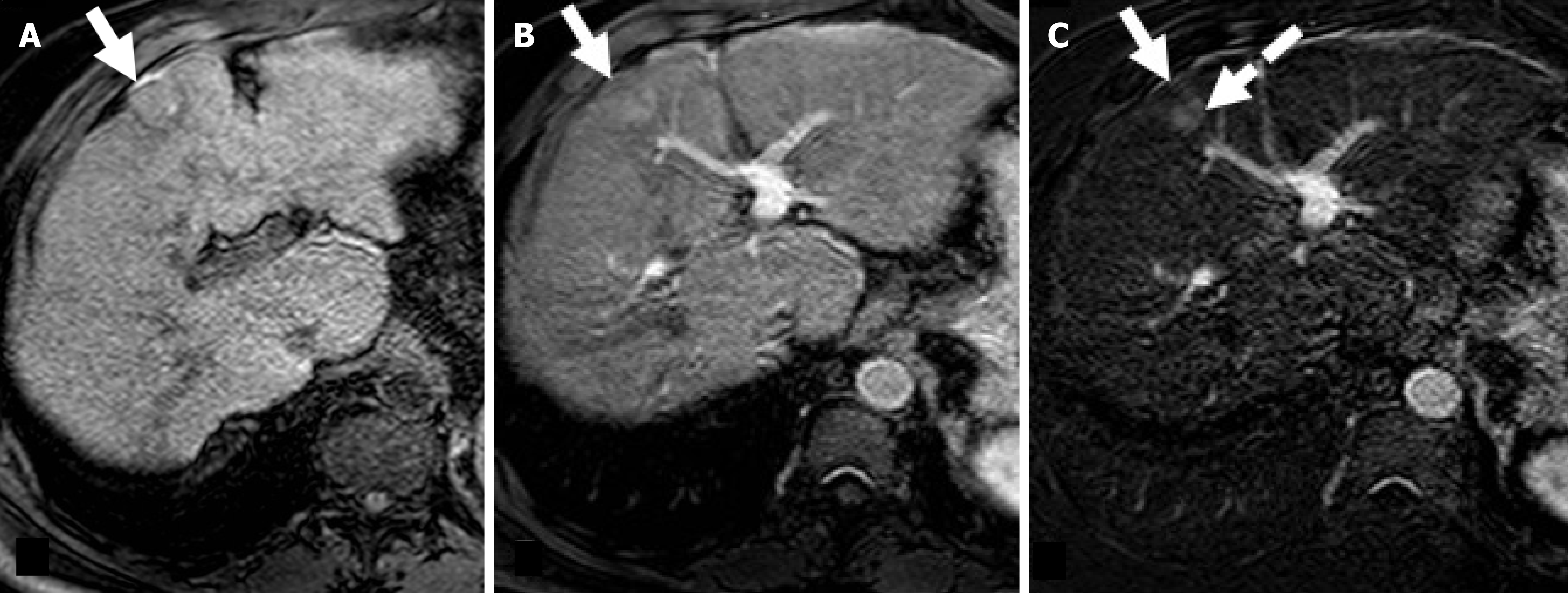
Figure 3 Residual hepatocellular carcinoma after radiofrequency ablation in a 68-year-old man with hepatitis C virus-related hepatic cirrhosis.
A: On magnetic resonance imaging obtained one month after radiofrequency ablation the treated area (arrow) shows slight hyperintensity on unenhanced T1-weighted due to coagulative necrosis; B: On corresponding hepatic arterial phase (HAP) image the treated area shows heterogenous hyperintensity; C: Subtracted HAP image shows enhancement in the posterior portion of the treated area (dotted arrow) suggesting residual tumor enhancement.
- Citation: Agnello F, Taibbi A, Galia M, Orlando A, Gagliardo C, Bartolotta TV. Hepatocellular carcinoma treatment response: Imaging findings and criteria. World J Radiol 2025; 17(10): 108804
- URL: https://www.wjgnet.com/1949-8470/full/v17/i10/108804.htm
- DOI: https://dx.doi.org/10.4329/wjr.v17.i10.108804