
















©The Author(s) 2025.
World J Radiol. Jan 28, 2025; 17(1): 97255
Published online Jan 28, 2025. doi: 10.4329/wjr.v17.i1.97255
Published online Jan 28, 2025. doi: 10.4329/wjr.v17.i1.97255
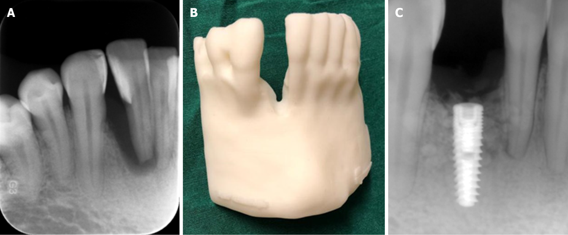
Figure 9 Cone beam CT images.
A-C: Intraoral periapical image (A) and three-dimensional printed bone model (B) of a patient from cone beam CT data obtained in order to assess right mandibular anterior lateral tooth for implant placement; C: According to diagnostic images and the three-dimensional bone model, an implant was successfully placed in the anterior lateral incisor region of the patient as can be seen from the postoperative periapical image.
- Citation: Kamburoğlu K. Trends in dentomaxillofacial radiology. World J Radiol 2025; 17(1): 97255
- URL: https://www.wjgnet.com/1949-8470/full/v17/i1/97255.htm
- DOI: https://dx.doi.org/10.4329/wjr.v17.i1.97255