
















©The Author(s) 2024.
World J Radiol. Sep 28, 2024; 16(9): 380-388
Published online Sep 28, 2024. doi: 10.4329/wjr.v16.i9.380
Published online Sep 28, 2024. doi: 10.4329/wjr.v16.i9.380
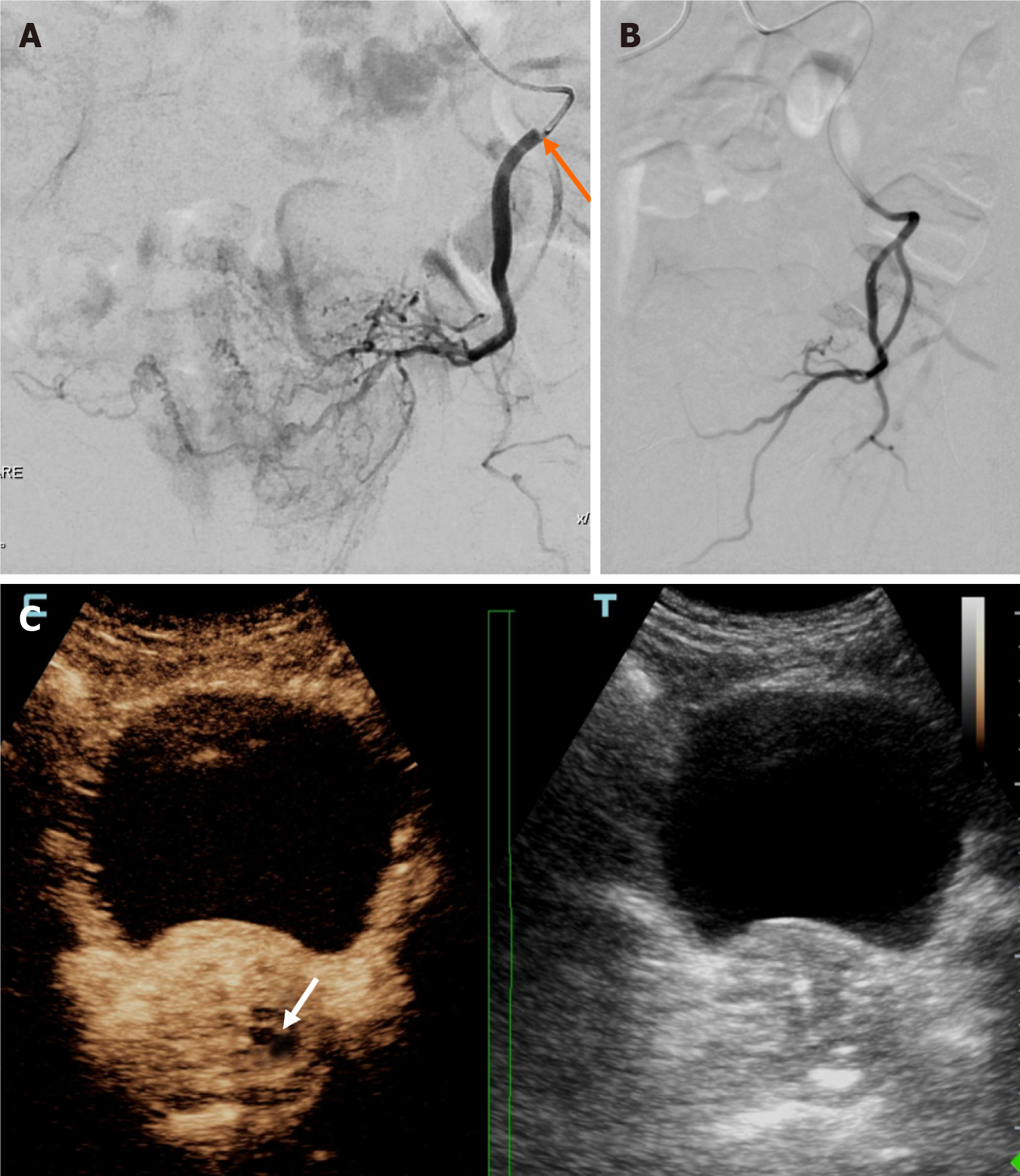
Figure 4 Representative images from a clinically unsuccessful case of intentionally unilateral prostatic artery embolization (subgroup A).
A: Left prostatic artery (PA) angiogram (frontal projection) with the microcatheter tip near the origin of the PA (arrow) shows opacification of the left lobe and of the largest part of the right lobe; B: Left PA angiogram (frontal projection) post embolization, from a more proximal position, shows apparent devascularization of the prostate; C: Dual image from intraprocedural contrast-enhanced ultrasonography (contrast-enhanced image on the left, unenhanced, reference B-mode image on the right) 2 minutes post embolization, shows minimal infarction (arrow) in the left lobe.
- Citation: Moschouris H, Stamatiou K. Intentionally unilateral prostatic artery embolization: Patient selection, technique and potential benefits. World J Radiol 2024; 16(9): 380-388
- URL: https://www.wjgnet.com/1949-8470/full/v16/i9/380.htm
- DOI: https://dx.doi.org/10.4329/wjr.v16.i9.380