
















©The Author(s) 2024.
World J Radiol. Aug 28, 2024; 16(8): 294-316
Published online Aug 28, 2024. doi: 10.4329/wjr.v16.i8.294
Published online Aug 28, 2024. doi: 10.4329/wjr.v16.i8.294
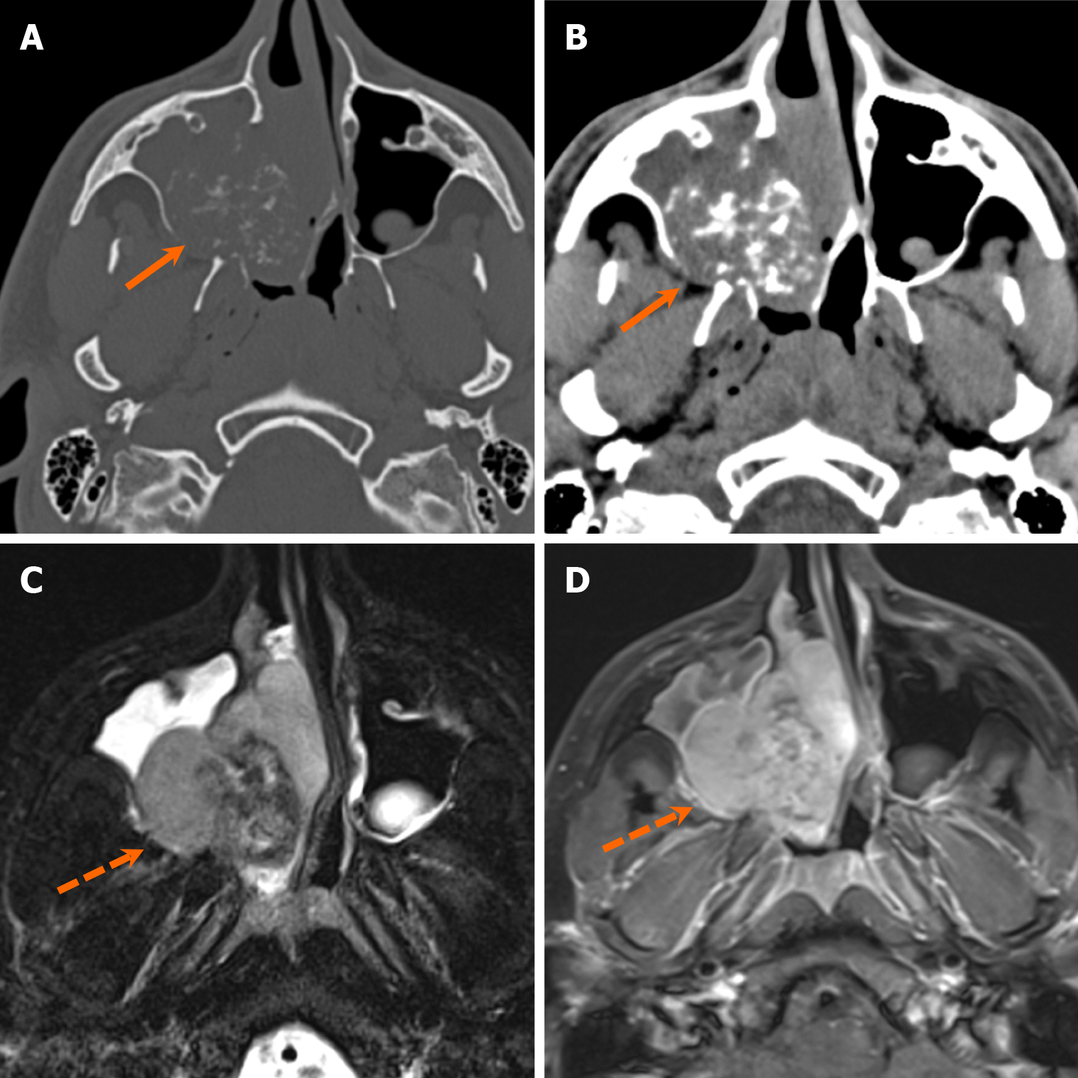
Figure 24 Chondrosarcoma.
A 19-year-old man with right nasal obstruction. A and B: Axial computed tomography images in bone (A) and soft tissue (B) windows depict an expansile right maxillary soft tissue mass with internal “ring-and-arc” calcifications (arrows); C and D: Axial T2-weighted (C) and contrast-enhanced fat-suppressed T1-weighted (D) magnetic resonance images demonstrate an enhancing mass (arrowheads) with heterogenous intermediate to low T2 signal due to internal calcifications. Pathology confirmed mesenchymal chondrosarcoma following resection.
- Citation: Choi WJ, Lee P, Thomas PC, Rath TJ, Mogensen MA, Dalley RW, Wangaryattawanich P. Imaging approach for jaw and maxillofacial bone tumors with updates from the 2022 World Health Organization classification. World J Radiol 2024; 16(8): 294-316
- URL: https://www.wjgnet.com/1949-8470/full/v16/i8/294.htm
- DOI: https://dx.doi.org/10.4329/wjr.v16.i8.294